Reference Range
Gram stain is the most common stain used in microbiology to identify bacteria. It is named after Christian Gram, who first developed the technique to identify the organism responsible for pneumonia.
A normal value is defined as no pathologic organism seen in the smear.
Normal findings: Normal upper respiratory tract [1]
Interpretation
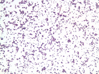
Gram-positive organisms are seen as blue/purple (see image below).
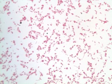
Gram-negative organisms are seen as pink/red (see image below).
Gram-variable organisms are those that cannot be grouped as either negative or positive.
The appearance of organisms that stain gram-positive or -negative means that the smear contains organisms. These organisms may be pathogenic or nonpathogenic. Further identification is required to interpret the results.
Collection and Panels
The specimen can be collected from any source (eg, sputum, wound, cerebrospinal fluid [CSF], pleural fluid, peritoneal fluid, urine, nose, rectum).
The specimen is spread on a slide and a thin smear should be prepared, as thick smears may diminish the accuracy of interpretation. There should be no areas with inconsistency or clumps.
Slides can also be made from cultured specimens or tissues.
The staining procedure consist of 7 steps, as follows [2] :
The smear is fixed with 95% methanol for 2 minutes or heat fixation technique
After preparing the smear, the slide is stained for one minute with primary stain, which consists of crystal violet
The slide is washed with flowing water for 2 seconds
The slide is flooded with Gram iodine, which acts as a mordant, for one minute
The slide is washed with flowing water for 2 seconds
A 50-50 mixture of acetone and 95% ethanol is used as decolorizing agent for 15 seconds
The slide is flooded with counterstain safranin for 30 seconds to 1 minute
After staining, the slide is examined under microscope with oil immersion. The organism is identified as gram-positive or -negative based on the color and as bacilli or cocci based on the shape. Bacilli are rod-shaped organisms, while cocci are spherical in shape.
A variation of Gram staining is called Atkin Gram stain, in which the primary stain is gentian violet and mordant is Atkin iodine. This mordant is more effective in retaining the primary stain. These make stain less susceptible to decolorization.
Background
Description
Gram-positive organisms have a higher content of peptidoglycan in their cell wall than gram-negative organisms. Approximately 40%-80% of the dry weight of gram-positive bacteria consists of peptidoglycan, while gram-negative bacteria have only a single layer of peptidoglycan. [3]
When cells are stained with crystal violet, crystal violet binds to the cell wall. When ethanol is used as decolorizing agent, gram-negative bacteria lose their outer membrane because of the higher content of lipid, allowing crystal violet complex to leak through the cell wall. In contrast, in gram-positive organisms, because of the higher content of peptidoglycan in the cell wall, crystal violet is trapped inside the cell. Thus, they stain a purple/blue color. Gram-negative organisms take the counterstain and stain pink/red.
Indications/Applications
Gram stain is used to identify the organism causing a disease or pathological condition.
Examples of gram-positive cocci include Staphylococcus and Streptococcus species.
Gram-positive bacilli include Corynebacterium, Clostridium, and Listeria species.
Gram-negative cocci include Neisseria gonorrhoeae, Neisseria meningitides, and Moraxella species.
Gram-negative bacilli include Escherichia coli, Pseudomonas species, Proteus species, and Klebsiella species.
Gram-variable organisms include Actinomyces species.
Considerations
The smear should be thin and not clumped.
The decolorizing time may differ based on the thickness of the smear; thin smears require shorter decolorizing time than thick smears.
If the organism culture is old, it may lose the peptidoglycan cell wall, causing a gram-positive organism to appear as gram-negative or gram-variable.
Organisms without a cell wall (eg, Mycoplasma species) and small bacteria (eg, Chlamydia and Rickettsia species) do not stain with Gram stain. [4, 5]
Legionella species do not stain but are visualized from growth in a culture medium.
Mycobacteria usually do not stain with Gram stain.
False results may result from the following circumstances:
-
Patient receiving antibiotics before specimen collection
-
Very young or very old culture
-
Inexperience
-
Excessive heat fixation
-
Improper decolorization
Enhancement methods to visualize gram-negative organisms include the following [6] :
-
Staining organism darker with methylene blue
-
Contrasting background from organisms with tartrazine fast green
-
Cytocentrifuge technique
KOH staining can be used to help differentiate gram-negative organisms from gram-positive ones based on the viscosity and string out of the suspension. [7, 8]
-
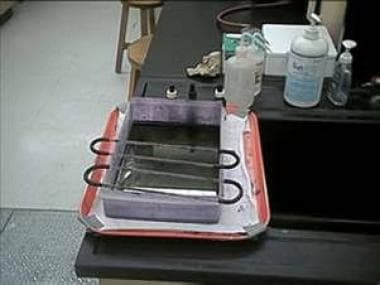
Tray for staining slides.
-
Gram-positive cocci.
-
Gram-negative bacilli.