History
Symptoms
Symptoms are as follows:
-
Painless skin patch. Chronic insensate patch is seen in the image below.
 Chronic insensate patch due to leprosy infection. Ho Chi Minh City, Vietnam. Courtesy of D. Scott Smith, MD.
-
Loss of sensation or paresthesias where the affected peripheral nerves are distributed
-
Wasting and muscle weakness
-
Foot drop or clawed hands (may result from neuritic pain and rapid peripheral nerve damage; as seen in the image below)
-
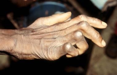 Characteristic clawed hand deformity caused by ulnar involvement in leprosy. Daloa, Ivory Coast. Courtesy of D. Scott Smith, MD.
-
Ulcerations on hands or feet (ulcer at the metatarsal head is seen in the image below)
-
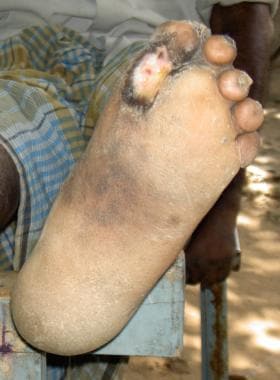 Chronic nonhealing ulcer at the metatarsal head resulting from loss of sensation in the feet. Karigiri, Tamil Nadu, India. Courtesy of Tara Ramachandra.


Ridley-Jopling classification [15]
Signs and symptoms of tuberculoid leprosy include the following:
-
Painless pale or red skin lesions with loss of sensation; lesions become raised as the disease progresses
-
A few affected nerves with diminished sensation and burning or tingling sensations
-
Significant sensory loss early in the disease course
-
Milder disease and fewer bacteria
Signs and symptoms of lepromatous leprosy include the following:
-
Painless pale or red skin lesions without loss of sensation; lesions become raised as the disease progresses
-
Thickening of peripheral nerves with diminished sensation and burning or tingling sensations
-
Extensive sensory loss over a longer period
-
More severe disease and larger numbers of bacteria
-
Facial deformities and paralysis
-
Involvement of eyes, bones, and other tissues
WHO Classification
The WHO Classification of leprosy is as follows:
-
Multibacillary: More skin lesions
-
Paucibacillary: Fewer skin lesions
-
Single-lesion paucibacillary: Single skin lesion
Symptoms in reactions
Symptoms in reactions are as follows:
-
Type 1 (reversal) - Sudden onset of skin redness and new lesions
-
Type 2 (erythema nodosum leprosum [ENL]; as seen in the image below) - Many skin nodules, fever, redness of eyes, muscle pain, and joint pain (seen in lepromatous leprosy only) [15]
-
Lucio phenomenon - Multiple ulcers
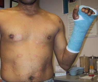 Patient with erythema nodosum leprosum type 2 reaction several weeks after initiation of drug therapy. This photograph was taken after tendon release. Redwood City, California. Courtesy of D. Scott Smith, MD.
Travel
Leprosy should be considered in anyone who has lived in the tropics or who has traveled for prolonged periods to endemic areas.
Exposure
The incubation period of leprosy is long, ranging from a few months to 20-50 years. The mean incubation time is estimated to be 10 years for lepromatous leprosy and 4 years for tuberculoid leprosy. The organism's slow dividing time (once every 2 wk) contributes to the challenge of epidemiologically linking exposures to the development of disease.
Because of immunologic reasons, only around 5-10% of the population is estimated to be susceptible to infection.
Physical
The cardinal signs of leprosy include hypoesthesia, skin lesions, and peripheral neuropathy. The first physical signs of leprosy usually are cutaneous. The subtype of leprosy often determines the degree of skin involvement.
Physical examination
Physical examination should include the following:
-
Evaluation of skin lesions
-
Careful sensory and motor examination
-
Palpation of peripheral nerves for pain or enlargement, with particular attention paid to the following locations:
Elbows - Ulnar nerve
Wrist - Superficial radial cutaneous and median nerves
Popliteal fossa - Common peroneal nerve
Neck - Great auricular nerve
Physical findings in specific leprosy subtypes
Tuberculoid leprosy
The initial lesion often is a sharply demarcated hypopigmented macule that is ovoid, circular, or serpiginous. The lesions may be somewhat elevated with a dry scaly center and erythematous borders.
Common lesion sites include the buttocks, face, and extensor surfaces of limbs. The perineum, scalp, and axilla are not normally involved because of the temperature differential in these zones, as predilection is toward cooler zones.
As the disease progresses, lesions tend to destroy the normal skin organs such as sweat glands and hair follicles.
Superficial nerves that lead from the lesions tend to enlarge and sometimes are palpable. The patient may experience severe neuropathic pain. Nerve involvement can lead to trauma and muscle atrophy.
Lepromatous leprosy
This form is characterized by extensive bilaterally symmetric cutaneous involvement, which can include macules, nodules, plaques, or papules. Multiple flat hypopigmented lesions are seen in the image below.
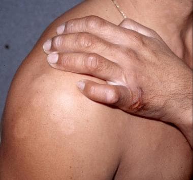 Multiple flat hypopigmented lesions on shoulder and neck, suggestive of multibacillary leprosy. Note ulceration of hypothenar area of hand, indicative of sensory loss with curled 5th digit, from ulnar neuropathy. Redwood City, California, United States. Courtesy of D. Scott Smith, MD.
Unlike lesions in tuberculoid leprosy, those in lepromatous leprosy have poorly defined borders and raised and indurated centers. As in all forms of leprosy, lepromatous lesions are worse on cooler parts of the body. Common areas of involvement include the face, ears, wrists, elbows, buttocks, and knees.
Hoarseness, loss of eyebrows and eyelashes, and nasal collapse secondary to septal perforation may occur in advanced cases of disease. Involvement of the eye may include keratitis, glaucoma, or iridocyclitis as seen in the image below.
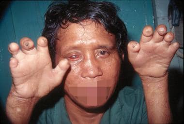 Man with advanced deformities caused by unmanaged leprosy. Keratitis, loss of eyebrow, thickened skin, and typical hand impairments. Ho Chi Minh City, Vietnam. Courtesy of D. Scott Smith, MD.
The leonine facies associated with leprosy develop as the disease progresses, and the facial skin becomes thickened and corrugated.
Axillary and inguinal adenopathy may develop, in addition to scarring of the testes and subsequent gynecomastia and sterility.
Nerve involvement in lepromatous leprosy is not as severe as in tuberculoid leprosy, since nerves, although visibly thickened and highly infected, still function reasonably well in early stages of the disease.
Borderline tuberculoid leprosy
The lesions are few or moderate and asymmetric with almost complete anesthesia. Peripheral nerves often are involved and thickened asymmetrically, and cutaneous nerves are sometimes enlarged.
Mid-borderline leprosy
The number of skin lesions is moderate, and they are asymmetrical and somewhat anesthetic. Peripheral nerves may be somewhat symmetrically enlarged, but cutaneous nerves are not.
Borderline lepromatous leprosy
Moderate to numerous slightly asymmetrical skin lesions appear with minor or no anesthesia. Peripheral nerves often are enlarged symmetrically, but cutaneous nerves are not.
Indeterminate leprosy
Skin lesions are typically either hypopigmented or hyperpigmented macules or plaques. Patients may note that these lesions are anesthetic or paresthetic.
Causes
M leprae is the causative agent associated with leprosy, which has been recognized as an infectious disease for the last 2 millennia. M leprae was discovered as the causative agent in 1873. The acid fast, gram-positive bacillus is an obligate intracellular organism with a predilection for Schwann cells and macrophages.
The route of transmission has not been definitively established, although human-to-human aerosol spread of nasal secretions is thought to be the most likely mode of transmission in most cases. Leprosy is not spread by touch, since the mycobacteria are incapable of crossing intact skin. Living near people with leprosy is associated with increased transmission. Among household contacts, the relative risk for leprosy is increased 8- to 10-fold in multibacillary and 2- to 4-fold in paucibacillary forms. Animal reservoirs do exist (armadillos, certain nonhuman primates), and cases of suspected zoonotic transmission have been reported.
-
Hands with Z-thumbs, clawing, contractures, and shortening of fingers due to repetitive injury and healing. Ho Chi Minh City, Vietnam. Courtesy of D. Scott Smith, MD.
-
Patient with facial nerve palsy and contractures of the hand. Daloa, Ivory Coast. Courtesy of D. Scott Smith, MD.
-
Chronic insensate patch due to leprosy infection. Ho Chi Minh City, Vietnam. Courtesy of D. Scott Smith, MD.
-
Characteristic clawed hand deformity caused by ulnar involvement in leprosy. Daloa, Ivory Coast. Courtesy of D. Scott Smith, MD.
-
Chronic nonhealing ulcer at the metatarsal head resulting from loss of sensation in the feet. Karigiri, Tamil Nadu, India. Courtesy of Tara Ramachandra.
-
Multiple flat hypopigmented lesions on shoulder and neck, suggestive of multibacillary leprosy. Note ulceration of hypothenar area of hand, indicative of sensory loss with curled 5th digit, from ulnar neuropathy. Redwood City, California, United States. Courtesy of D. Scott Smith, MD.
-
Man with advanced deformities caused by unmanaged leprosy. Keratitis, loss of eyebrow, thickened skin, and typical hand impairments. Ho Chi Minh City, Vietnam. Courtesy of D. Scott Smith, MD.
-
Histopathology of leprosy: Large numbers of acid-fast bacilli (in clusters) in histiocytes and within nerves. Fite-Faraco stain 500 X. Courtesy of Tara Ramachandra, MD, and D. Scott Smith, MD.
-
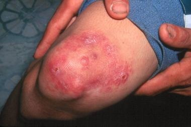
Patient with multibacillary leprosy showing subsequent erythema nodosum leprosum reaction. Santa Clara, California. Courtesy of D. Scott Smith, MD.
-
Patient with erythema nodosum leprosum type 2 reaction several weeks after initiation of drug therapy. This photograph was taken after tendon release. Redwood City, California. Courtesy of D. Scott Smith, MD.
-
Increased pigmentation on the face due to clofazimine therapy. Courtesy of D. Scott Smith, MD.
-
2018 leprosy treatment guidelines. Courtesy of the WHO.
-
Map of countries reporting rifampicin resistance in leprosy between 2009 and 2015. Courtesy of the WHO.
-
2018 WHO guidelines for treatment of drug-resistant leprosy. Courtesy of the WHO.
-
2018 guidelines for single-dose rifampicin. Courtesy of the WHO.
-
WHO map showing new cases of new leprosy cases, 2020. Courtesy of the WHO.
Tables
Age Group |
Drug |
Dosage and Frequency |
Duration |
|
Multibacillary Leprosy |
Paucibacillary Leprosy |
|||
Adult |
Rifampicin |
600 mg once a month |
12 months |
6 months |
300 mg once a month and 50 mg daily |
||||
100 mg daily |
||||
Children (10-14 years) |
Rifampicin |
450 mg once a month |
12 months |
6 months |
Clofazimine |
150 mg once a month, 50 mg on alternate days |
|||
Dapsone |
50 mg daily |
|||
Children < 10 years or < 40 kg |
Rifampicin |
10 mg/kg once a month |
12 months |
6 months |
Clofazimine |
100 mg once a month, 50 mg twice weekly |
|||
Dapsone |
2 mg/kg daily |
|||
Resistance Type |
Treatment |
|
First 6 months (daily) |
Next 18 months (daily) |
|
Rifampicin resistance |
Ofloxacin 400 mg* plus Minocycline 100 mg plus Clofazimine 50 mg |
Ofloxacin 400 mg* or Minocycline 100 mg plus Clofazimine 50 mg |
Ofloxacin 400 mg* plus Clarithromycin 500 mg plus Clofazimine 50 mg |
Ofloxacin 400 mg* plus Clofazimine 50 mg |
|
Rifampicin and ofloxacin resistance |
Clarithromycin 500 mg plus Minocycline 100 mg plus Clofazimine 50 mg |
Clarithromycin 500 mg or Minocycline 100 mg plus Clofazimine 50 mg |
*Ofloxacin 400 mg can be replaced by levofloxacin 500 mg or moxifloxacin 400 mg |
||


