Background
The laboratory exercise provides instruction on the techniques needed to perform a differential Gram stain. Bacteria can be divided into two groups: gram positive and gram negative. The type of cell wall of a bacteria determines its gram type. If the cell wall consists of a thick peptidogycan layer then the bacteria is gram-positive. If the bacterial cell wall consists of a thinner peptidoglycan layer surrounded by a lipid layer then the bacteria is gram-negative (Cappuccino 75).
The results of a gram stain can help in determining the course of treatment for a patient. The gram stain procedure can be performed on body fluids, throat cultures, and sputum specimens to determine the gram type of the organisms present. This information can then be used to decide what additional tests to perform to identify the strains of bacteria present (Cappuccino 77).
Materials and Methods
The following materials were used:
Nutrient agar slant cultures (E. coli and S. aureus)
Reagents (crystal violet, Gram’s iodine, 95% ethyl alcohol, and safranin)
Bunsen burner
Inoculating loop
Staining tray
Glass slides
Bibulous paper
Lens covers
Microscope
The following procedure was followed:
To begin the experiment, eight clean glass slides were obtained. Then, using aseptic technique, a smear of S. aureus, E. coli, and a mixture of the two specimens was prepared. To prepare the slides, a drop of water on each individual slide, and then each organism was transferred separately to the drop of water with a sterile, cooled loop. To sterilize the loop, the coil was placed through the blame of a Bunsen burner until it turned red, and then allowed to cool. The necks of the test tubes containing the cultures were flamed as well to prevent cross-contamination. Both organisms were mixed and spread by means of a circular motion of the cooled inoculating loop. The slides were labeled A (S. aureus), B (E. coli), and C (Mixture).

(Three slides, post-staining)

(Crystal violet, Gram’s iodine, and safranin)
To begin the gram staining, drops of crystal violet were gently flooded on the slides, and allowed to rest for one minute. The crystal violet stain was used to stain all cells purple. After one minute elapsed, the slides were gently washed with tap water. It was important to not allow the stain to sit for too long, as it could have resulted in over-staining.
Following the rinse, the slides were flooded with Gram’s iodine, allowed to sit for one minute, and then gently rinsed with tap water. Gram’s iodine served not only as a killing agent, but as a mordant as well. A mordant is a substance that increases a cell’s affinity for a stain. It binds to the primary stain and forms an insoluble complex. The resultant complex serves to intensify the color of the stain.
Following the rinse of the Gram’s iodine, the slides were decolorized with 95% ethyl alcohol. The ethyl alcohol served as a protein-dehydrating agent and as a lipid solvent. It was important not to over-decolorize the smears. The ethyl alcohol was slowly added drop by drop until the alcohol ran almost clear and showed a blue tinge.
The slides were rinsed again, and then counterstained with safranin for 45 seconds. Safranin is used to stain pink the cells that were decolorized.
The slides were rinsed once more and then blotted dry with bibulous paper. The three slides were finally examined under oil immersion.
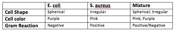
Results
Review Questions:
1. What are the advantages of differential staining procedures over the simple staining technique?
Differential staining procedures can help identify the presence of different organisms in a culture specimen. Some of the organisms will stain with the primary stain color and others with the counterstain color.
2. Cite the purpose of each of the following reagents in a differential staining procedure.
a. Primary stain. The primary stain is used to stain all cells in the sample
b. Mordant. The mordant is used to increase the cell’s affinity for the stain. It binds the primary stain and forms an insoluble complex.
c. Decolorizing agent. The decolorizing agent is used to remove the color from the cells that have a thinner peptidoglycan cell wall.
d. Counterstain. The counterstain is used to stain those cells that have been previously decolorized.
3. Why is it essential that the primary stain and the counterstain be of contrasting colors?
The primary stain and counterstain need to be of contrasting color to help visualize the number cells that retained the primary stain.
4. Which is the most critical step in the performance of the Gram staining procedures? Explain.
The most critical step in the performance of the Gram staining procedure is the application of the decolorizing agent. If too much decolorizing agent is applied, the crystal violet will was out from gram positive cells.
5. Because of a snowstorm, your regular laboratory session was canceled and the Gram staining procedure was performed on cultures incubated for a longer period of time. Examination of the stained B. cereus slides revealed a great deal of color variability, ranging from an intense blue to shades of pink. Account for this result.
In cultures that are older than 24 hours, the gram positive organisms may lose their ability to retain the primary stain. Some of the gram positive cells may appear purple and others purple.
Works Cited
Cappuccino, James G. Microbiology: a laboratory manual – 10th ed. Glenview, IL: Pearson, 2014. Print.
Last updated on 7-April-2014 at 2:41 PM
