History
Common historical features of rectus sheath hematoma (RSH) include acute abdominal pain, fever, nausea, and vomiting. The nonspecific nature of these symptoms combined with the low incidence of the disorder lead to difficulty in considering this diagnosis. Rectus sheath hematoma should be included in the differential diagnosis of every patient who presents with abdominal pain.
Specific symptoms
Constitutional
Fever and chills are common symptoms in rectus sheath hematoma. Symptoms of hypovolemic shock with weakness, confusion, pallor, and diaphoresis can develop in patients with a large rectus sheath hematoma.
Abdominal pain
The most common presenting symptom is acute abdominal pain. The onset of pain may be sudden, but more often, it develops over a period of several hours. The pain is typically sharp and severe, with an associated palpable abdominal mass. Pain is usually worse with movement and is often unilateral. Constant pain with episodic abdominal cramping is also a frequent symptom. In atypical cases, the pain may develop insidiously, making the abdominal mass difficult to differentiate from an abdominal wall neoplasm.
Gastrointestinal/urologic
Anorexia, nausea, vomiting, diarrhea, constipation, tenesmus, and bladder irritability are all compatible with the diagnosis of rectus sheath hematoma. The severity of symptoms is related to the degree of peritoneal irritation.
Precipitating factors
The clinician needs to have rectus sheath hematoma in the differential, or the diagnosis will be easily overlooked. A careful history should include directed questions regarding surgical procedures, occult blunt trauma, coughing, sneezing, constipation (straining at the stool), or exercise. In patients with certain medical problems, questions about recent asthma exacerbations, bronchitis, or upper respiratory tract infections may prove helpful. Rectus sheath hematoma must always be considered in abdominal pain patients on anticoagulants.
Physical Examination
Vital signs
A low-grade fever is common in rectus sheath hematoma. The hematoma can be large enough to compromise intravascular volume, with resultant signs of hypovolemic shock including hypotension, tachycardia, and tachypnea.
Abdominal examination
Typically, the abdominal examination reveals a palpable, painful, firm, nonpulsatile abdominal mass corresponding to the rectus sheath. The mass may be bilobar with a central groove. The mass does not move with respiration. Because the hematoma is deep to the subcutaneous tissue and rectus muscles, the mass is not always palpable, particularly in obese patients. In 2000, Berna et al's case series reported a palpable mass detected in 8 of 12 patients. [20]
Hyperesthesia of the overlying skin is not uncommon. Bowel sounds may be absent. Signs of local peritoneal irritation with rebound tenderness and involuntary guarding may be present. This finding is most often seen in infra-umbilical hematomas due to the thin transverse fascialis serving as the only barrier between a hematoma and the peritoneum. Rarely, a hematoma may cause extraperitoneal compression of the abdominal cavity and cause abdominal compartment syndrome, or even rupture into the peritoneum, causing a chemical peritonitis.
The Fothergill sign is useful in determining whether an abdominal mass is part of the abdominal wall or whether it is in the abdomen. [21] It is elicited by voluntary contraction of the rectus muscles by the patient lifting either his or her head or legs while in the supine position. With this action, rectus sheath hematomas become fixed, more painful, and more tender, while intra-abdominal masses become less distinct and less tender. The Fothergill sign may be inconclusive in patients who are obese or pregnant. As described by Fothergill in 1926 [22] :
This patient complains of pain and the medical man finds the swelling. The trouble is that he seldom knows how long the swelling has been present…The main point is the recognition that these swellings are part and parcel of the abdominal wall. This is generally made by noting that they can still be felt when the recti are in action, and that they become fixed as the muscles contract
The Carnett sign is an additional test to assist in differentiating between abdominal wall and intra-abdominal pathology. [21] It is performed by having the patient lie supine and tensing the abdominal musculature by raising either the head or the shoulder off the table. A positive sign is elicited if abdominal tenderness is increased or unchanged while tensing the abdomen. This indicates an abdominal wall process. A negative sign, or decreased abdominal tenderness while tensing the abdomen, suggests intra-abdominal pathology. Previous studies have demonstrated this sign to be fairly sensitive but not specific for abdominal wall pathology.
The Cullen sign of periumbilical ecchymosis is associated with retroperitoneal or abdominal wall hemorrhage. In rectus sheath hematoma, ecchymosis appears after 2-5 days. The ecchymosis uncommonly extends into the flanks.
See the image below.
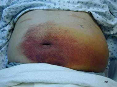 Rectus Sheath Hematoma. The Cullen sign, periumbilical ecchymosis, in a patient with a rectus sheath hematoma.
The Grey-Turner sign is another manifestation of retroperitoneal hemorrhage. This finding of flank ecchymosis was initially described in hemorrhagic pancreatitis, and along with the Cullen sign, it is not specific for retroperitoneal or abdominal wall hemorrhage.
Pelvic examination
The pelvic examination may reveal a mass anterior to the vagina and above the pubis. The pelvic examination may be misleading, particularly in those cases that demonstrate unilateral adnexal tenderness and mass.
-
Rectus Sheath Hematoma. Anatomy of the rectus sheath.
-
Rectus Sheath Hematoma. The Cullen sign, periumbilical ecchymosis, in a patient with a rectus sheath hematoma.
-
Rectus Sheath Hematoma. Rectus sheath hematoma of the right rectus muscle. Image courtesy of Dr David Gordon.
-
Rectus Sheath Hematoma. Note how the rectus sheath hematoma becomes bilobar as it dissects inferiorly (same patient as in the previous image). Image courtesy of Dr David Gordon.
-
Rectus Sheath Hematoma. Ultrasound image of a rectus sheath hematoma presenting as a tender, unilateral abdominal mass. Source: Maharaj D, Ramdass M, Teelucksingh S, Perry A, Naraynsingh V. Rectus sheath haematoma: a new set of diagnostic features. Postgraduate Medical Journal. 2002;78:755-6. Reproduced with permission from the BMJ Publishing Group.


