Request a callback
Contact us
Get Direction
Zehra Manzil, G-3, 91, Lady Jamshedji Rd, near Paradise cinema, Mahim West, Mumbai, Maharashtra 400016
What is Ultrasound?
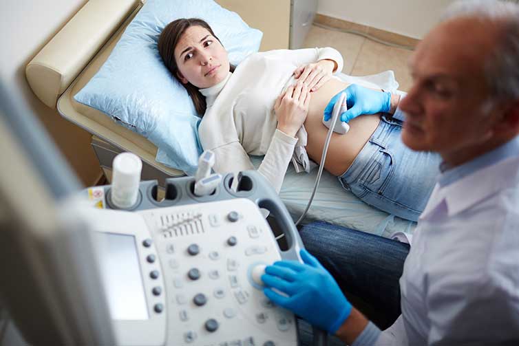
Ultrasound, also known as sonography, ultrasonography, USG, is a non-invasive, radiation free mode of diagnostic imaging technique used to assess the various abdominal and pelvic organs, vascular structure, muscles and tendons and also to evaluate the congenital abnormalities and well-being and optimal growth of foetus during pregnancy.
Ultrasound for pregnancy or USG for pregnancy is a primary mode of diagnostic imaging technique to provide diagnosis.
With the advances in ultrasound, we can obtain 3D ultrasound/4D ultrasound images of the foetus, Demarcate the lesions in thyroid and breast by elastography and also know the architecture of the liver using shear wave technique.
Why is Ultrasound Test/Sonography Test performed?
- To diagnose pathologies of abdomen like liver/ spleen enlargement, appendicitis, bowel obstruction, various hernias.
- To diagnose pathologies of kidneys such as calculi, cysts, mass lesions, urinary tract infection.
- To diagnose pathologies of pelvis in males like prostatomegaly (BPH).
- To diagnose pathologies of pelvis in females like fibroids, ovarian cysts, PCOD.
- For follicular monitoring.
- To diagnose pregnancy, know number of foetuses, detection of congenital foetal abnormalities, Colour Doppler study to evaluate foetal blood flow.
- Doppler ultrasound studies of arteries and veins to see for varicose veins, peripheral vascular disease.
- Renal Doppler, Carotid Doppler and Portal Doppler.
- Scrotal Doppler for evaluation of varicocele.
Is it safe?
Ultrasonography test is protected, noninvasive, and doesn’t use any radiation. Sonography tests are carried out on the surface of the skin, and there are no known complications associated with sonography test.
At Shanti Diagnostics we offer the following ultrasonography tests:
1. USG Abdomen and Pelvis:
A detailed whole abdomen ultrasound and pelvis ultrasound to look for pathologies of liver, pancreas, gallbladder, spleen, both kidneys, urinary bladder, prostate in male while uterus and ovaries in the females.
2. USG KUB (Kidney-ureter-bladder):
A detailed ultrasound to look for pathologies of both kidneys, ureters, urinary bladder, and prostate in male.
3. USG Pelvis:
A detailed pelvis ultrasound to look for pathologies of urinary bladder, prostate in male while uterus, ovaries and adnexa in the females.
4. USG Scrotum (Testes):
A detailed ultrasound to look for pathologies of both testes, epididymis, inguinal region and also to look for varicocele.
5. USG Breast (Sono-Mammography):
A detailed ultrasound to look for pathologies of bilateral breasts, nipple-areola complexes and bilateral axillae.
6. USG Colour Doppler of Peripheral limbs (Arterial Doppler / Venous Doppler):
A detailed ultrasound to look for arterial or venous insufficiencies of peripheral limb vessels, varicose veins, deep venous thrombosis, PIVD, arterial occlusion, diabetic neuropathy.
7. USG B-Scan:
A detailed ultrasound to look for pathologies of the orbit such as cataract, vitreous hemorrhage, retinal detachment and foreign body impaction.
8. USG Neck:
A detailed ultrasound to look for pathologies of the thyroid gland, submandibular and parotid glands and also lymph node associated pathologies.
9. USG Chest:
A detailed ultrasound of the chest to look for pleural pathologies, diaphragmatic movements and look for pneumothorax.
10. USG Inguinal Region:
A detailed ultrasound of the inguinal region to look for hernia, lymphadenopathy or other mass lesions.
How is the Ultrasound Test or Sonography Test conducted?
A disposable bedsheet is placed on a sanitized bed.
The patient is usually in supine position with part to be examined exposed. A special water-based gel heated to room temperature is applied to investigate the sound vibrations with a transducer. The radiologist uses the transducer across the part to be examined. The gel assists in getting perspectives on the desired tissues, and constructions. The gel is later wiped off after the test.
The entire procedure takes approximately 15 minutes to 1 hour, depending on the test. Once the test is complete, the high-quality images derived, printed on a glossy paper are used to analyse the results, which are later handed to the patient.
What are the preparations required for Ultrasonography Test?
Scans such of whole abdomen ultrasound, kidneys require fasting of about 4-6hrs. Other special tests require specific preparations as needed. The patient is always advised to carry previous reports for comparison.