The 20 best science images of the year?
- Published

From multicoloured scans of parts of the human body to vivid photos of creatures up close, the finalists of the annual Wellcome Image Awards have been announced.
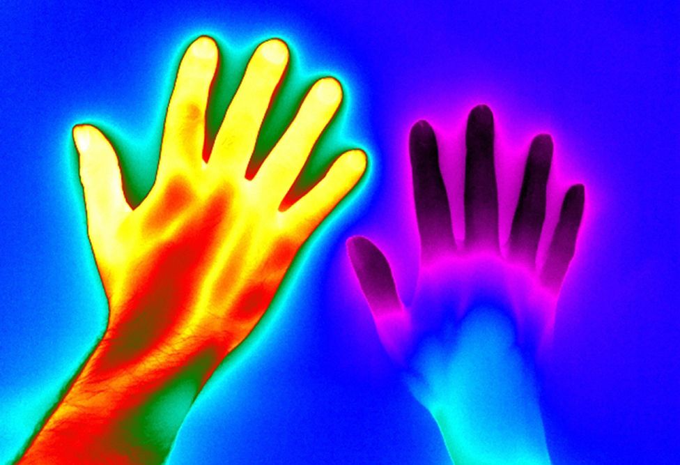
The thermal image above shows the temperature of two people's hands - a healthy person on the left, and someone with Raynaud's disease on the right.
Both hands were put in cold water for two minutes before being imaged. The healthy hand then warmed at a considerably faster rate.
"This image is striking because it shows so vividly the difference between normal circulation and the poor circulation of someone with Raynaud's disease - triggered by cold temperature, stress and anxiety," says head of Wellcome Images and chair of the judging panel, Catherine Draycott.
Scroll down to see the other 19 finalists.
Wiring the human brain
Alfred Anwander, Max Planck Institute for Human Cognitive and Brain Sciences
A kaleidoscope of colour reveals a map of pathways inside the brain of a young healthy adult.
Different parts of the brain communicate with each other through nerve fibres - which are colour-coded here.
Using a type of magnetic resonance imaging - or MRI - the image was created from mapping virtual slices of the brain, from top to bottom, tracking the direction and movement of water molecules.
"We felt it captured the essence of the technique whilst giving a picture of the living brain," says Catherine Draycott.
Black henna allergy
Nicola Kelley, Cardiff and Vale University Hospital NHS Trust
"Here you can see a black henna tattoo on the forearm of a young woman who has suffered an allergic reaction to the dye," explains Dirk Pilat, medical director for e-Learning at the Royal College of GPs, and a GP himself.
"It's beautifully lit and shows the translucence of the skin that's been raised in blisters, capturing the early stage of the reaction."
Dye from the henna plant is commonly used to temporarily stain skin or hair orange-brown, but chemical dyes can be added to turn the colour black.
Human stem cell
Sílvia A Ferreira, Cristina Lopo and Eileen Gentleman, King's College London
"This is a scanning electron micrograph of a stem cell taken from the bone marrow inside the hip bone of a healthy person," explains Robin Lovell-Badge, a Wellcome awards judge and head of stem cell biology and developmental genetics at the Francis Crick Institute.
"This really stood out, we found the natural symmetry alongside the very subtle colouring very striking. It's lovely and sharp."
Stem cells can divide to make some of the other types of cells found in the body.
This one is about 15 micrometres (0.015mm) across - and before the image could be taken, it was first frozen at cryogenic temperatures (lower than −150C or −238F).
Dividing stem cell in the brain
Paula Alexandre, University College London
This swirling pattern shows different stages of a stem cell splitting in two inside the brain of a zebrafish before it hatches.
The circle is about 250 micrometres (0.25mm) wide, and covers a time period of nine hours.
Starting at about the eight o'clock position, the cell splits to make two different cells found in the brain.
"It helps us visualise embryonic development by showing nature's beautifully orchestrated process of stem cell division - producing a new (purple) stem cell and a differentiated (white) nerve cell," says Anne Deconinck, executive director of the Koch Institute for Integrative Cancer Research at MIT, in the US.
Ebola virus
David S Goodsell, RCSB Protein Data Bank
On 15 March 2016 this image was named as the overall winner of the Wellcome Image Awards.
"This illustration shows the internal structure of an Ebola virus particle, with the central core in three dimensions so you can see the internal structures more clearly," says visual artist Rob Kesseler, professor at Central Saint Martins at the University of the Arts London.
The Ebola virus is about 100 nanometres (0.0001mm) wide - 200 times smaller than many of the cells that it infects.
"The illustrator chooses pastel colours rather than tonal contrast to show the different elements of this tiny, often lethal virus.
"It shows how illustration can uniquely show many different levels of detail simultaneously, with great beauty and clarity."
Infectious disease containment unit
David Bishop, Royal Free Hospital, London
"This photograph provides a rare glimpse inside the UKs only high-level isolation unit, taken the day before a nurse was admitted after contracting Ebola," says judge Rob Kesseler.
"It perfectly captures the calm before the storm, ghostly empty shapes of protective clothing hang, waiting for the patient to be rushed in."
This special see-through tent surrounds a bed in the Royal Free Hospital in London. All air leaving the unit is cleaned, so the patient can be safely treated without putting other patients or staff at risk.
Nurse Will Pooley made a full recovery.
Swallowtail butterfly
Daniel Saftner, Macroscopic Solutions
"It was chosen because it shows so clearly what the mouthparts and the eyes of a swallowtail butterfly look like in really striking detail," says judge Eric Hilaire, science, environment and global development online picture editor at the Guardian.
Butterflies have two big round eyes for seeing quick movements and two antennae for smelling.
They also have a long feeding tube, which is curled up like a spring here, but it unrolls so the butterfly can use it like a straw to drink nectar from flowers.
Moth scales
Mark R Smith, Macroscopic Solutions
"The colour here is actually an optical illusion," explains judge Eric Hilaire.
"The scales themselves don't contain much pigment, it's the way light bounces off the curves which gives them their apparent colour."
The scales belong to a Madagascan sunset moth - which sparkles with colour in the light and is often mistaken for a butterfly.
This picture is 750 micrometres (0.75mm) wide.
Inside the human eye
Peter Maloca, University of Basel
"Here we're looking inside blood vessels at the back of the human eye, which also supply the retina," says judge Robin Lovell-Badge of this 3D picture.
"The blood itself is moving too fast to be visible creating this maze of tunnels that looks like a subterranean landscape. It draws your eye towards what appears to be a light at the end."
Pictures like this are used by doctors to help spot early signs of eye disease. These tiny tunnels are about 100 micrometres (0.1mm) tall.
Blood vessels in the eye
Kim Baxter, Cambridge University Hospitals NHS Foundation Trust
"What's fascinating about this is that, when you see it, you don't automatically think of the eye," says judge Rob Kesseler. "It appears like an aerial view of a city at night or a telescopic image of a distant galaxy."
The image was created by photographing the blood vessels in the retina - seen here as white spidery lines - as fluorescent dye was passed through.
Detecting stroke
Nicholas Evans, University of Cambridge
"This demonstrates beautifully one of the deadliest two centimetres of pathology in human medicine and is an excellent tool to explain the causation of cerebrovascular accidents, or strokes, to patients," explains Dirk Pilat.
This medical scan shows, in green, a blocked blood vessel inside the neck of a person.
This vessel carries blood to the brain and when it gets blocked, parts of the brain can get damaged and stop working properly.
Cow heart
Michael Frank, Royal Veterinary College
"This image is striking in its three dimensional sculptural appearance, especially when you know that the heart itself is actually a specimen in a jar," says Catherine Draycott.
"Beautifully lit and photographed to bring to life an old historical specimen - highlighting both the external surface and internal structures."
Windows have been cut into this cow's heart to show what is inside. It is about four times the size of a human heart.
Engineering human liver tissue
Chelsea Fortin, Kelly Stevens and Sangeeta Bhatia, Koch Institute, MIT
This small piece of human liver has been put into a mouse with a damaged liver. The human liver has started to grow, with help from the mouse's blood.
"It is tissue engineering in action," says Anne Deconinck from the Koch Institute.
"In response to tissue damage, cells can reorganise and heal, and even develop much-needed blood vessels.
"This image with the heart-shaped patch of engineered liver cells beautifully conveys a message of hope - and the promise of scientific advancements to overcome the challenges of replacement organ shortages and disease, including cirrhosis and liver cancer."
This image appears as a result of the partnership between Wellcome Images and the Koch Institute at MIT, Cambridge, USA.
Bacteria on graphene oxide
Izzat Suffian, Kuo-Ching Mei, Houmam Kafa and Khuloud T Al-Jamal, King's College London
"This image serves really well to illustrate the fact that graphene, a recently discovered material, is just one atom thick," says Catherine Draycott.
"The details of the creases in the graphene contrast with the enormous torpedo shaped bacteria, which we know to be very small organisms."
Graphene - seen here in purple - is an extremely thin sheet of carbon, and is one of the thinnest, strongest materials so far discovered.
Researchers are trying to stick different medicines to it so they can be carried to the right place in the body when needed. The bacteria are about two micrometres (0.002mm) long.
Premature baby receiving light therapy
David Bishop, Royal Free Hospital, London
On 15 March 2016, at the Wellcome Image Awards, this image was named as the winner of the Julie Dorrington Award for outstanding photography in a clinical environment.
This baby was born early and has jaundice, a common condition which turns the skin and eyes yellow.
The baby is being treated in an incubator at Barnet Hospital in north London, and lies under a blue coloured light, with eyes covered.
"One of the reasons this was picked is that it is intimate yet respectful - due to the framing and angle of the photograph," says Catherine Draycott.
Clathrin cage
Maria Voigt, RCSB Protein Data Bank
"Clathrin is a protein found in cells, and here molecules of it have come together to form this cage like structure which helps move things around the cell," explains judge Eric Hilaire.
"The illustration and its shading bring out the three dimensional nature of this structure."
Cells can have lots of these tiny cages inside them. This cage measures about 50 nanometres (0.00005mm) across.
When the cage is not being used it breaks up into smaller pieces, which get recycled. The cage can be put back together again when it's next needed.
Toxoplasmosis-causing parasites
Leandro Lemgruber, University of Glasgow
"It looks quite blurry because of the extreme magnification of this tiny parasite which causes toxoplasmosis," says judge Robin Lovell-Badge.
"This infinitesimally small organism, found in infected cat faeces and raw or undercooked meat, is particularly dangerous to pregnant women and people with weakened immune systems."
Here, DNA inside each parasite (blue/green) is surrounded by membrane (red) and protein (black).
This image was created using a type of super-resolution microscopy. Each parasite measures 10 micrometres (0.01mm) long.
Bone development
Frank Acquaah
"Here modern technology is used on historical remains," says Catherine Draycott. "The images are made with micro-computed tomography - penetrating wave scans - which show how the internal structure of the bone evolves as a baby develops in the womb and after birth."
Each circle shows bone from an infant at a different age. The youngest (three months before birth) is on the left and the oldest (2.5 years old) is on the right. These historical bones all come from the skeletons of children who died in the 19th Century.
Maize leaves
Fernan Federici, Pontificia Universidad Catolica de Chile and University of Cambridge
"This image evokes the work of Gustav Klimt with its beautiful, gilded mosaic look," says Anne Deconinck from the Koch Institute.
This is a confocal micrograph looking inside a cluster of leaves from a young maize (corn) plant.
Each curled leaf is made up of lots of small cells (small green square and rectangle shapes) - and inside each cell is a nucleus (orange circle), the part of the cell which stores genetic information.
The overall winner of the Wellcome Image Awards will be announced on the evening of Tuesday 15 March 2016 at the Science Museum in London - where all the images will be on display until Sunday 19 June 2016.
All images subject to copyright - courtesy Wellcome Images.