Diagnostic Sonography
Date: July 13, 2017
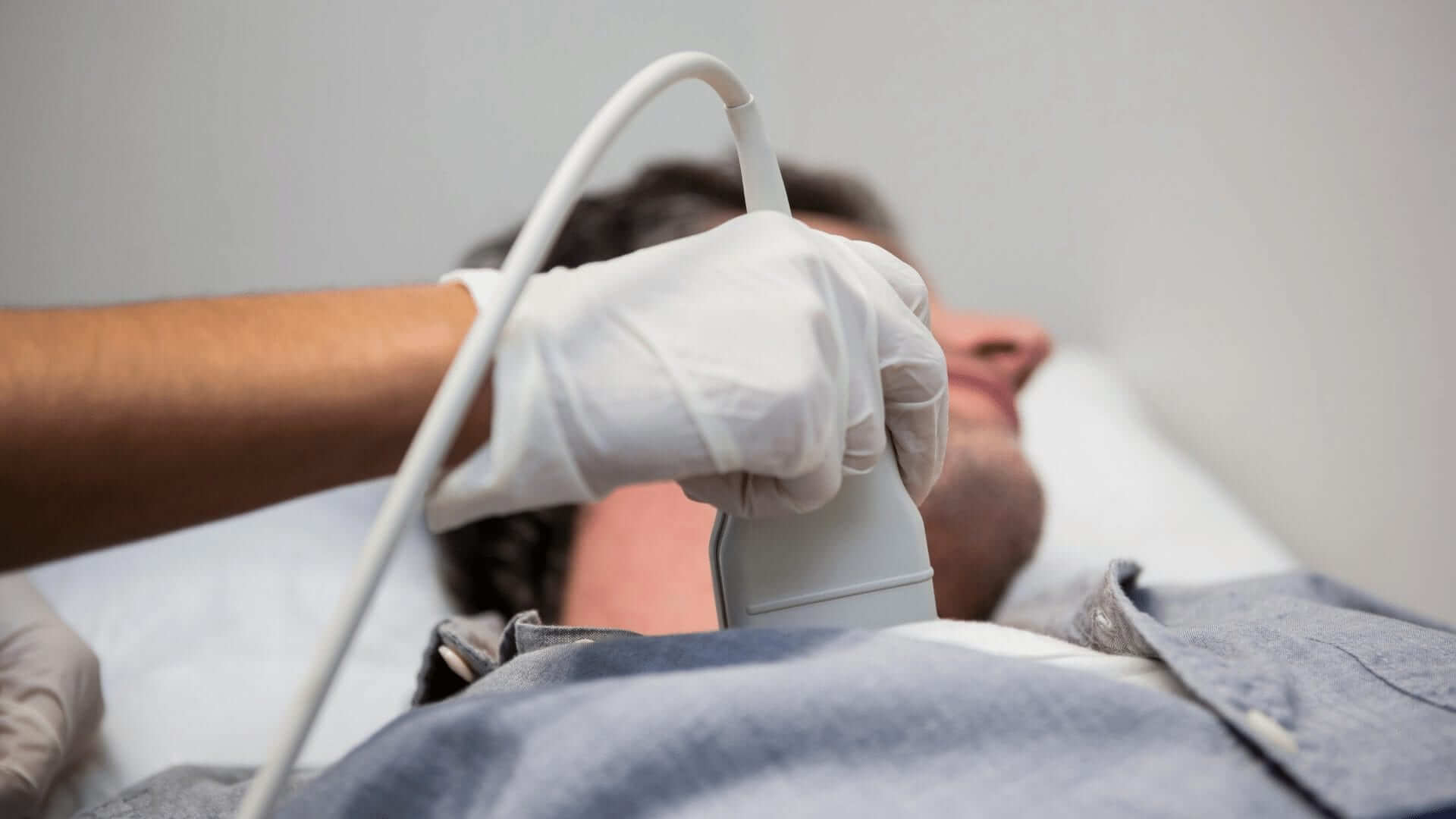
Diagnostic sonography is a medical procedure based on high-frequency sound waves used to obtain images of organs, tissues, blood vessels, and blood flow to diagnose an ailment. Ultrasound scan and sonogram are terms referring to the same procedure. Some of the body parts that may be examined using this method are the abdomen, breasts, heart, and circulatory system.
More and more, this procedure is used to detect and treat heart disease, heart attack, and disease affecting blood vessels that may determine a stroke. Diagnostic medical sonography also proves to help do tissue biopsy. The advantage of this imaging procedure is that no forms of radiation are involved. Those professionals who perform diagnostic medical sonography but are not physicians are called sonographers or ultrasound technicians.
There are various specialty areas in the field of diagnostic medical sonography. For starters, investigating the abdomen involves evaluating soft tissue, blood vessels, and organs in the abdomen. Sonographers may diagnose or treat conditions involving kidneys, liver, gallbladder, bile ducts, spleen, and pancreas. Breast abnormalities will be evaluated with this method after they have been discovered with screening or diagnostic mammography.

Diagnostic Medical Sonography
Breast sonography may be used to track tumors, blood supply conditions, and biopsies of breast tissue. Diagnostic medical sonography is also used in obstetrics/gynecology. The female reproductive system can be investigated with this procedure. When it comes to pregnant women, their babies’ growth may be tracked, and their health kept under observation.
Cardiovascular sonography is meant to evaluate blood vessels, blood flow, the heart, and its valves. Neurosonology is used to investigate the brain and the spinal cord. Neurological and nervous system disorders may be diagnosed in premature infants that are in neonatal care. Diagnostic medical sonography may also offer images of the eye and its muscles.