
Nanotechnology in Food and Plant Science: Challenges and Future Prospects


Department of Epidemic Disease Research, Institute for Research and Medical Consultations (IRMC), Imam Abdulrahman Bin Faisal University, P.O. Box 1982, Dammam 31441, Saudi Arabia
Plants 2023, 12(13), 2565; https://doi.org/10.3390/plants12132565
Submission received: 6 June 2023
/
Revised: 24 June 2023
/
Accepted: 29 June 2023
/
Published: 6 July 2023
(This article belongs to the Special Issue Nanotechnology in Plant Science)
Abstract
:Globally, food safety and security are receiving a lot of attention to ensure a steady supply of nutrient-rich and safe food. Nanotechnology is used in a wide range of technical processes, including the development of new materials and the enhancement of food safety and security. Nanomaterials are used to improve the protective effects of food and help detect microbial contamination, hazardous chemicals, and pesticides. Nanosensors are used to detect pathogens and allergens in food. Food processing is enhanced further by nanocapsulation, which allows for the delivery of bioactive compounds, increases food bioavailability, and extends food shelf life. Various forms of nanomaterials have been developed to improve food safety and enhance agricultural productivity, including nanometals, nanorods, nanofilms, nanotubes, nanofibers, nanolayers, and nanosheets. Such materials are used for developing nanofertilizers, nanopesticides, and nanomaterials to induce plant growth, genome modification, and transgene expression in plants. Nanomaterials have antimicrobial properties, promote plants’ innate immunity, and act as delivery agents for active ingredients. Nanocomposites offer good acid-resistance capabilities, effective recyclability, significant thermostability, and enhanced storage stability. Nanomaterials have been extensively used for the targeted delivery and release of genes and proteins into plant cells. In this review article, we discuss the role of nanotechnology in food safety and security. Furthermore, we include a partial literature survey on the use of nanotechnology in food packaging, food safety, food preservation using smart nanocarriers, the detection of food-borne pathogens and allergens using nanosensors, and crop growth and yield improvement; however, extensive research on nanotechnology is warranted.
1. Introduction
The economic strength of a country mainly depends on agriculture and its products. Improved agricultural practices, including soil preparation and irrigation, are necessary to achieve self-sufficiency in food production and to strengthen a country. Self-sufficiency may be gained by solving agricultural problems, protecting food, and improving farmers’ knowledge about using modern agricultural technologies [1]. Globally, food safety and security are currently receiving a great deal of attention with the aim of ensuring a sustainable supply of nutrient-rich and safe food. Specifically, the Kingdom of Saudi Arabia is among the largest countries, with approximately 30 million citizens; it has been reported that about 80% of its food demand is met via importing food from other countries, which clearly poses a significant challenge to the economy, food safety, and public health [2]. Moreover, food can also be spoiled by various foodborne pathogens or chemical contaminants, which may cause diseases and sometimes the death of consumers. Thus, to ensure food safety, food handling, distribution, storage, and food quality should be improved; such measures would not only enhance food consumption but also prevent negative effects on consumers’ health [3].
Recently, nanotechnology has been used for a broad range of technical processes, including the characterization, fabrication, and regulation of various materials, in order to develop new materials and to create chemical, biological, biomedical, pharmaceutical, agricultural, electronic, and bioengineering applications [4,5,6,7,8,9,10,11,12,13]. Nanotechnology deals with the manipulation of materials (molecules or macromolecules), devices, or systems at the nanometer scale in order to create and utilize materials with unique properties. The developed nanomaterials are 1–100 nm in size and possess a high surface-to-volume ratio; they have unique physiochemical properties, including color, strength, diffusion, and toxicity [14]. Due to these unique properties, nanomaterials are also used to improve the protective qualities of food. Additionally, in the detection of microbial contamination, hazardous chemicals, and pesticides, various nanosensors and nanopackaging materials have been used to increase sensitivity and specificity. Food processing is further improved by nanocapsulation, which enables the delivery of bioactive compounds, increases the bioavailability of food, and improves the shelf life of foodstuffs [15]. Various forms of nanomaterials have been developed to improve food safety and enhance agricultural productivity, including nanorods, nanofilms, nanotubes, nanofibers, nanolayers, and nanosheets. In this study, we analyzed and highlighted the importance of the application of nanotechnology in improving food safety, security, processing, and packaging; we also address its relation to food insecurity issues, whereby nanotechnology is used to solve agricultural problems, such as post-harvest losses and crop growth and yields. Furthermore, we discuss the application of nanotechnology in biotic/abiotic stress tolerance and the detection of food allergens and food-borne pathogens using nanobiosensors to improve food productivity and quality. Additionally, we discuss the limitations, rules and regulations, challenges, and future prospects of using nanotechnology in the food industry and related industries.
2. Food Security and Safety
Food security refers to all people globally having physical and economic access to sufficient, safe, and nutritious food that meets their dietary needs, allowing them to live active and healthy lives. A lack of food security leads to chronic diseases due to malnutrition. The four most important pillars of food security are food availability, food stability, food utilization, and food access [16,17]. According to the United Nations Food and Agriculture Organization (UNFAO), the foremost universal challenge that is likely to appear by 2050 is an increase in food demand, ranging from 59 to 98%, due to the rapidly rising global population, particularly in developing countries, which is expected to reach 9 billion [18]. To ensure food security, a 70% increase in food production is required, which will be affected by climate change, land availability, agricultural yields, and food supply. In addition, the UNFAO noted that, while food waste occurs in rich countries due to packaging expiration and aesthetic concerns, it occurs more frequently in developing countries due to spoiling, contamination, and poor food quality [19,20]. Abiotic stresses (such as the salinization of the soil, elevated CO2 levels and temperatures, drought, and nutritional imbalances) have been shown to negatively affect the development and growth of plants and their productivity and quality. In certain cases, these factors have even led to the local extinction of some species. Moreover, abiotic stresses have reportedly caused an average yield loss of more than 50% in the majority of crops [21,22]. Therefore, to reduce the agricultural chemical footprint, it is essential to enhance plants’ growth and tolerance to abiotic and/or biotic stresses, agricultural productivity, and, most importantly, their quality through the application of nanotechnology and bioactive compounds found in seaweeds/plants [21]. Food waste also affects the environment by impacting land and water availability for food production and increasing greenhouse gases that lead to climate change [23].
Food safety is another major public health concern related to protecting food from physical, chemical, and biological contaminants during food preparation, processing, handling, and distribution, thereby protecting consumers [24,25]. Food safety is significantly affected by changes in food recipes, food habits, toxins, pathogens, and contaminants, which cause serious threats to people’s health [24]. In a country like Saudi Arabia, there is limited agricultural productivity and, thereby, completely relies on food imports and observance of Islamic laws; thus, food security is highly challenging for national control and regulatory institutions [26]. Farmers throughout the world are looking for innovations and new technologies that help to preserve food safety and security by enhancing crop production, soil improvement, food packaging, distribution, and food quality [27]. Conventional methods for the analysis of contaminations, pathogen detection, and improving food security are time consuming and expensive. Thus, recently, in the developing world, the use of many nanoscale materials and their devices has been developed with low-cost and large- areas for improving agricultural efficiency, soil improvement, food processing and packaging, food shelf-life, and the analysis of pathogens and their toxins [23].
3. Nanotechnology in Food Security and Safety
A technology that increases production while decreasing food waste is required to ensure global food security. Recently, nanotechnology has provided a path for increasing food availability and developing new products that are beneficial to agriculture, particularly by minimizing nutrient losses in fertilizers and increasing food yield through pest and nutrient management, among other things [28]. It can also be used to improve food processing, create novel food products with enhanced tastes and flavors, extend the shelf life of food products, and develop nano-based methods for detecting and preventing food spoilage (Figure 1) [29].
3.1. Nanotechnology Application in Crop Growth and Yield
Nanotechnology is used to address various agricultural issues by developing nanofertilizers, nanopesticides, and nanomaterials to induce plant growth, genome modification, and transgene expression in plants [30,31]. In a study, a chitosan nanofertilizer containing copper and salicylic acid was developed and tested for its source activity in maize plants. Seed treatment with chitosan nanofertilizer showed a significant increase in seedling vigor index and increased reserve food mobilizing enzyme activities, while foliar application suggested that used for seed treatment and foliar application of chitosan nanofertilizer significantly increased antioxidant enzymes, decreased lipid peroxidation, stimulated chlorophyll content, and ultimately suggested that nanofertilizer, by slowly releasing copper and salicylic acid, effectively enhanced crop yield by promoting source activity [32]. El-Naggar et al. [33] developed silica NPs (SiO2-NPs) and applied them as nanofertilizers and pesticides to maize plants. Based on the plant height, chlorophyll content, and protein composition, the data showed that minerals (nitrogen, phosphorus, and potassium) interacted with nanoparticles and could be used as growth promoters and pesticides for crops during storage at a very low and safe dose. The nanopowder waste-derived NPK fertilizers were produced from squid pin waste, shrimp shells, bovine bone, banana peels, and Kolakhar, and foliarly applied Capsicum annum L. cv. Cordoba plants were equated with commercial chemical fertilizers. The data showed that the nanocomposite increased plant growth and yield compared to commercial fertilizer-treated plants, suggesting that waste nano-NPK fertilizer can be used as an alternative to foliar fertilizer to increase Capsicum production [34]. Silver nanoparticles (AgNPs) at a dose of 15 mL/L increased the chlorophyll content, fruit yield, stem diameter, and flower percentages compared to Zinc NPs (ZnO NPs) at a dose of 7.5 mL/L. Additionally, the pollen size and spores were also enhanced by spraying AgNPs. Moreover, AgNPs-treated peach leaf extracts exerted antibacterial activity at a dose of 3000 µg/mL with a zone of inhibition in the range of 6–8.67 mm [35]. In a study, Moringa oleifera tree growth in calcareous soil was treated with vermicompost and NPK (NPs) fertilizer in combination for two seasons. The data showed that pod and seed yields were significantly increased, and the fixed oil of the tree was also improved by the combination of vermicompost and nanoparticles under calcareous soil conditions [36]. A study was conducted by applying 20–40 nm iron (III) oxide (Fe2O3) nanomaterials (NMs) in a hydroponic system to wheat plants. It has been reported that it increased plant height, chlorophyll content, root length, and iron content of wheat without any toxicity compared to 30–50 and 8–10 nm Fe2O3 NMs, ultimately suggesting that it could be an effective nanofertilizer for agricultural applications [37]. The effect of Fe2O3NPs (γ-Fe2O3-maghemite and Fe3O4-magnetite) was tested on barley plants, and it was reported that Fe2O3 NPs enhanced the germination rate, barley biomass, and pigmentation, thereby improving plant growth. It was also suggested that iron oxide nanoparticles could be used as nanofertilizers [38]. Recently, a novel phosphorus nanofertilizer was synthesized using hydroxyapatite NPs, pomegranate peel, and coffee ground extracts, and it was reported that the green synthesized nanofertilizer modulated photosynthetic potential, levels of carbohydrates, metabolites, and biocompatibility, and also decreased cytotoxic and genotoxic parameters in Punica granatum L. [39]. The effects of the three iron forms, namely, nano (Fe-NPs), sulfate (FeSO4), and chelated (Fe-chelated), were investigated by foliar spraying on Washington navel orange trees, and it was reported that the Fe-chelated and Fe nano forms improved vegetative growth, flower set, tree nutritional status, enhanced fruit production, and increased the quality of plant growth in environmentally preferred arid regions [40].
Nanotechnology is a new research arena, chiefly in using nanopesticides made up of NPs to control disease in plants, and is gaining attention nowadays. Bacterial panicle blight disease affects rice yield worldwide due to seed-borne rice pathogens like Burkholderia glumae and B. gladioli. A study was conducted on the biosynthesis of ZnO NPs from a native Bacillus cereus RNT6 strain against B. glumae and B. gladioli. The data revealed that NPs exerted antibacterial potential against B. gladioli and B. glumae by increasing the zone of inhibition, decreasing cell numbers, and inducing morphological damage, and were suggested as effective nanopesticides to act upon bacterial panicle blight disease [41]. The application of metal oxide NPs like CuO, ZnO, and FeO reduced bacterial wilt incidence and also decreased the rhizosphere bacterial community by affecting its composition and structure. Among all the metal oxide nanoparticles, CuO NPs effectively reduced the abundance of pathogens and suggested that metal oxide NPs can be used for controlling and preventing tomato bacterial wilt, a soil-borne disease [42]. A study has developed a nanopesticide formulation using chitosan and a botanical pesticide, PONNEEM®, which showed effective antifeedant activity and larvicidal potential against Helicoverpa armigera [43]. Green-synthesized CdS NPs were tested against malaria vectors and showed high toxicity in young mosquitoes, Anopheles stephensi and A. sundaicus, as well as impacting the acetylcholinesterase and glutathione S-transferase activities of the mud crab Scylla serrata [44].
Recently, nanotechnology has helped to distribute genetic materials to plants to perform genetic engineering and stabilize genetic materials for plant improvement [45]. In a study, it was reported that carbon dots (NPs) can act as carriers to deliver the plasmids into mature plants to develop transient plant transformation. A foliar application of plasmid-coated carbon dots on wheat plants is used to deliver cas9, gRNA, and GFP, suggesting that spray-on gene editing is simple, inexpensive, rapid, and transforms in plants [46]. In another study, double-stranded RNA that could induce RNA interference (RNAi) was sprayed on plants to modify traits without inducing genetic modification. For this, double hydroxide layered NPs with a diameter size of 50 nm were used to deliver dsRNA effectively into tomato pollen with an incubation of 2–4 h, which led to a decrease in the level of miRNA with early bicellular pollen 3 d post-treatment, thus ultimately suggesting that layered double hydroxide NPs could be used as a promising nonviral vector for other biomolecules into plant cells and effective delivery of dsRNA [47]. Carbon nanotubes with a single-walled chitosan-complex were developed using the penetration mechanism of a lipid exchange envelope into chloroplasts for delivering plasmid DNA of various plant species, including Spinacia oleracea, Nasturtium officinale, and Nicotiana tabacum, without the help of any chemical or biological methods. It acts as an effective transgene delivery tool compared to conventional delivery methods for the benefit of plant growth [48].
3.2. Application of Nanotechnology in Controlling Plant Diseases
Plant pathogenic bacteria, viruses, and fungi cause devastating diseases in hundreds of crops worldwide, leading to agricultural losses by affecting crop yield, quality, and growth. Thus, to counteract plant and post-harvest diseases, it is necessary to develop novel techniques and tools to bypass conventional agriculture practices. Climate change will affect crop production by increasing plant diseases and reducing the efficacy of disease-control approaches. Table 1 lists the roles of nanomaterials in plant diseases. However, nanotechnology, particularly the use of nanomaterials, effectively involves plant disease management using the following three mechanisms: (1) acting as antimicrobials; (2) acting as stimulants to induce the innate immunity of plants; and (3) acting as delivery agents for active ingredients, including pesticides and micronutrients [49]. Nanotechnology provides a solution for disease management by providing efficient formulations for the delivery of agrochemicals, nutrients, and growth regulators for the management of plant diseases that could be pathogens, thereby preventing crop loss [50]. To decrease phytotoxicity, nanocapsules improve herbicide application, enhance penetration through cuticles and tissues, and allow a slow and steady release of active ingredients inside the plant. NPs act like ‘magic bullets’, loaded with chemicals or nucleic acids, and target specific plant areas to release their charge [51]. Tomato bacterial wilt is one of the soil-borne bacterial infectious diseases caused by Ralstonia solanacearum. The structure and composition of rhizosphere bacterial colonies were reduced by synthesized metal oxide nanoparticles like CuO, ZnO, and FeO. Comparatively, CuO NPs significantly reduced pathogens and enhanced physiological parameters in tomato bacterial wilt-infected tomato plants, suggesting that metal oxide NPs can be effectively used against soil-borne bacterial pathogens [42,52]. In a study, TiO2, Ag, ZnO, and SiO2 NPs were synthesized with sizes of 25, 11, 35, and 25 nm and tested against a model plant-associated bacterium, Pseudomonas protegens CHA0, at 250 μg/mL. At a sublethal dose (500 ng/mL), NPs stimulated the antifungal effect of P. protegens by stimulating prm operon expression, which would enhance the synthesis of the antifungal molecule pyrrolnitrin [53]. Thus, NPs application could be an effective measure to excite the antifungal activity of pseudomonads and could be used as apromising disease control agent.
Ginger essential oil is composed of monoterpenes and sesquiterpene hydrocarbons, which can effectively affect the cell structure and internal structure of pathogens and inhibit various phytopathogens. However, the solubility and stability of compounds in ginger essential oil are one of the important drawbacks, and they can be fixed by using nanoemulsions to increase the potential and delivery of essential oils against various pathogens to control plant diseases [54]. In a study, dsRNA was loaded onto layered double hydroxide (LDH) clay nanosheets, which are non-toxic, degradable, and show sustained release upon spraying on plant leaves. It remains in the plant for at least 30 days post-application and offers plant protection against viruses for about 20 days [55]. The protective effect of MgO NPs was tested against plant diseases using root dip, soil drench, and foliar methods and showed that MgO exerted a twin role by acting as a nematocidal and growth-promotion inducer at a concentration of 100 ppm by decreasing nematode fecundity, altering the size of galls, stimulating pigments like chlorophyll and carotenoids, enhancing protein and nitrogen content at the root and shoot, and inducing plant growth [56]. In a study, four types of copper NPs (types 1 to 4) were tested against Fusicladium oleagineum and Colletotrichum spp., which cause olive leaf spots in the olive plant. Comparatively, CuNP Type 1 and Type 2 effectively reduced mycelial growth, whereas Type 3 and Type 4 exerted strong protective effects against both species [57]. In another study, synthesized chitosan-coupled copper NPs were tested against damping-off fungal diseases in plant seeds. Chitosan-coupled copper NPs exert fungicide action by inhibiting mycelial growth and act as growth promoters by increasing growth-promoting activity in plants. It could be used as a safe alternative to commercially available pesticides by decreasing hazardous effects on the environment [58]. Nanocomposites were synthesized using copper oxide NPs decorated on the surface of reduced graphene oxide nanosheets to test against Fusarium oxysporum, which affects tomato and pepper plants. At 1 mg/L, the nanocomposite showed higher antifungal potential compared to fungicide Kocide 2000 (2.5 g/L) by increasing pits and pores on the fungal cell membranes, leading to cell death and thereby reducing the severity of Fusarium wilt and root rot disease. Ultimately, it was suggested that the synthesized nanocomposite could be an eco-friendly and economical nanopesticide for plant protection [59]. Green synthesized ZnO NPs using leaf extracts of Withania coagulans were tested for their antibacterial effect against the bacterial wilt pathogen in tomato plants. The combined treatment for 12 days of application time effectively suppressed Ralstonia solanacearum by decreasing disease severity and increasing plant growth [60]. Activation of ZnO NPs using visible light (405 nm, 18–30 J/cm2) inactivates the human pathogen E. coli and the wheat pathogen F. oxysporum without regrowth. The inactivation could be due to reactive oxygen species without affecting the visual quality of grains, preventing inhibition of germination rate, and enhancing grain seed growth, which would meet the increasing demand of the world population [61].

Table 1.
Application of nanotechnology in the prevention and control of plant diseases.
| S.No | Nanomaterials | Disease Causing Agent | Disease | Mechanism | Reference |
|---|---|---|---|---|---|
| 1. | AgNPs | Fusarium spp. (F. moniliforme and F. oxysporum) | Damping-off of cotton seedlings. | Fungal growth was reduced by Ag NPs (25 to 200 ppm) interaction | [62] |
| 2. | ZnO NPs | Tomato mosaic virus | Viral Infection in Tomato in Egypt | ZnO-NPs showed remarkably increased growth indices, photosynthetic attributes, and enzymatic and non-enzymatic antioxidants compared to the challenge control (interestingly, oxidative damage caused by ToMV was reduced by reducing malondialdehyde, H2O2, and O2 levels) | [63] |
| 3. | Bacillus-mediated synthesized AgNPs | S. viridans and R. solani | Sheath blight disease | Destruction of mycelia of R. solani by reducing lesion length and enhancing root and shoot length in Oryza sativa seeds. | [64] |
| 4. | AgNPs synthesized from peels of pomegranate and orange fruits extracts | Alternaria solani. | Early Blight Disease of Tomato in Saudi Arabia | Reduced the mycelial growth of A. solani compared to the crude extracts of two peels | [65] |
| 5. | Copper NPs using extracts of eucalyptus and mint leaves. | Colletotrichum capsici | Anthracnose of chili | 1000 ppm showed the highest mycelial inhibition of 99.78% | [66] |
| 6. | ZnO NPs | Fusarium oxysporum | Wilt disease in Solanum melongena L. | Improved plant height and root length Increased content of chlorophyll a and chlorophyll b Enhanced antioxidant activity and isozymes compared to infected control | [67] |
| 7. | ZnO NPs using Trichoderma harzianum | Fusarium sp., Rhizoctonia solani and Macrophomina phaseolina. | Fungal disease control in cotton | Reduced mycelial growth | [68] |
| 8. | Chitosan/dextran NPs | Alfalfa mosaic virus | Viral infection in Nicotiana glutinosa plants | Induced resistance, enhanced carbohydrates, and phenolic contents Increased expression of peroxidase and phenylalanine ammonia-lyase | [69] |
| 9. | Silicon dioxide NPs using saffron extract | Rhizoctonia solani | Damping-off and root rot diseases | Reduced mycelial radial growth Increased photosynthetic pigments (chlorophylls and carotenoids) Decreased oxidative stress by activating SOD, APX, CAT, phenolics, and flavonoids | [70] |
| 10. | Biosynthesized selenium NPs using B. megaterium | Rhizoctonia solani | Rhizoctonia root-rot disease | Improve morphological and metabolic indicators Minimize the severity of root rot disease | [71] |
| 11. | Gold NPs synthesized from Tinospora cordifolia | Pseudomonas aeruginosa | Biofilm-related infections | Reduced number of biofilm-forming cell internalization of the NPs, probably affecting the tendency of P. aeruginosa to colonize on the surface of the NPs | [72] |
3.3. Application of Nanotechnology in Improving Stress Tolerance
Salinity is an abiotic stress that has destructive effects on plants by affecting their growth, development, and yield, including rapeseed. Selenium and ZnO NPs were synthesized using plant hormones to study the effects of the germination process under salt stress. NPs reduced abscisic acid and regulated gibberellic acid, which promoted early seedling growth and seed germination under salt stress in rapeseed plants [73]. A carbon quantum dot nanoparticle is functionalized with proline to evaluate its effect on grapevine plants under salt stress. The NPs are reported to increase leaf fresh and dry weight; enhance superoxide dismutase, glutathione peroxidase, and catalase activities; enhance carotenoid, phenol, and anthocyanin content; and decrease malondialdehyde and hydrogen peroxide levels in plants under salinity (Figure 2). Ultimately, carbon quantum dots with chemical priming agents showed their plant priming effect by decreasing abiotic stress-related damage in plants [74]. Yttrium doping-stabilized γ-Fe2O3 NPs were supplied in nutrient solutions to Brassica napus plants, and it was found that NPs reduced H2O2 and lipid peroxidation, increased chlorophyll content, and improved plant growth under drought stress. Thus, maghemite NPs could be effectively used as plant fertilizer and help in drought stress management when compared to chelated iron [75]. Arbuscular mycorrhizal fungi (AMF) are symbionts that assist plants in overcoming environmental stress. Nanotechnology was used to retain AMF activity, for which TiO2 NPs were tested for the activity of AMF in bean roots under salinity stress, suggesting that TiO2 NPs showed positive effects confined to AMF under salinity stress and not on bean plants [76].
Synthesized chitosan–selenium NPs were foliar sprayed on bitter melon (Momordica charantia) under salinity stress, and it was reported that NPs increased antioxidant enzyme activity, proline, water, and potassium, and decreased malondialdehyde, hydrogen peroxide, and sodium aggregation in plant tissues, thereby enhancing salinity tolerance [77]. Foliar application of chitosan NPs improved banana plant growth, maintained the nutrient content of plants, increased the fresh and dry weights, stimulated photosynthetic pigments, decreased MDA, reduced the accumulation of free radical species (H2O2, •OH, O2•), enhanced the accumulation of osmoprotectants (soluble carbohydrates, proline, and amino acids), and ultimately developed cold stress tolerance [78].
Fluoride pollution stimulated bioaccumulation in roots and shoots that inhibited reproduction, agronomic development, and uptake of minerals and decreased enzymatic antioxidants, and enhanced reactive oxygen species-mediated oxidative injury. Banerjee and Roychoudhury [79] reported that nanomaghemite decreased fluoride accumulation, stimulated the uptake of potassium, calcium, iron, copper, zinc, and selenium, and decreased the translocation of cobalt. It also triggered plant growth; stimulated antioxidant enzymes like SOD, CAT, and GPX; enhanced proline, anthocyanins, and flavonoids; and ultimately helped plants develop the potential to scavenge free radicals. Thus, it was reported that iron nanotechnology is involved in stimulating safe rice cultivation in fluoride-polluted environments. Similarly, magnetite NPs help in decreasing the salt stress effects by increasing proteins, chlorophyll content, SOD, and glutathione in two wheat cultivars (Misr1 and Gimmeza11) and reducing the lipid peroxidation marker MDA content [80]. In another study, green-synthesized gold NPs were reported to modulate the K+/Na+ ratio, chlorophyll content, defense systems, nitrogen intake and output, stomatal responses, and oxidative stress in wheat plants under high salt stress [81]. Cerium oxide NPs enhanced plant photosynthesis, modified the anatomy of plant roots, and enhanced plant salt stress tolerance by forming apoplastic barriers in brassica roots, which allows more Na+ transport to shoots and less accumulation of Na+ in plant roots [82].
3.4. Application of Nanotechnology in Post-Harvest Loss Reduction
Worldwide, greater than 40% of food losses, including vegetables, cereals, fruit, fish, meat, and milk products, occur due to post-harvest, food processing, trade, and customer stages in both developed and developing countries. Zeolitic imidazole framework-8 (ZIF-8) nanocomposites were synthesized with tunable pore sizes and high surface areas and functionalized with catalase by controlled self-assembly of silanes or tannic acid and Fe3+. Nanocomposites are reported to be an effective strategy that possesses high acid resistance capacity, effective recyclability, high thermostability, and enhanced storage stability [83]. Certain bacteria are necessary for the improvement of soil quality and the enhancement of plant nutrient acquisition. In a study, nanocoating was synthesized with N-hydroxysuccinimide-modified poly (γ-glutamic acid) and calcium ions to attach to the surface of bacteria (P. stutzeri NRCB010) via the formation of covalent and ionic bonds on the surface of the bacteria. Nanocoating enhanced the bacteria’s resistance to harsh environments compared to uncoated bacteria and enhanced the shelf life of bacteria [84].
A photocatalyst, TiO2 was reported to possess excellent bactericidal potential under UVA light. Three methods, namely, the glass cover-slip, indented coupon, and direct spreading, were used to determine the bactericidal potential of TiO2, and among them, indented coupon techniques were more effective in determining the bactericidal potential of TiO2 [85]. Hannon et al. [86] reported that copper and AgNPs with polystyrene-polyethylene oxide block copolymer (PS-b-PEO) are used for coating food packages. Comparatively, silver coating lowered the migration of nanoparticles to humans compared to copper. However, silver and copper nanoparticles are safe for use in food packaging, but the margin of exposure to humans exceeds regulatory limits, so it is suggested that they should be confined to nanopackaging applications. A nanocoating agent, chitosan NPs, was developed using sodium tripolyphosphate penta basic via the ionic gelation method to extend the mean life of fresh-cut bell peppers by preventing microbial contamination with Listeria monocytogenes and Salmonella enterica. Nanocoating protected fresh-cut bell peppers for 12 days at a temperature of 5 °C without affecting weight or sensory quality [87]. In another study, Staphylococcus aureus, other microflora, and Pseudomonas fluorescens were isolated from raw chicken and tested against low-density polyethylene films with a silver coat. Additionally, chicken breast fillets were also packed with low-density polyethylene films with a silver coat using skin packaging with a vacuum upon storage. Nanocoating extends the shelf life of chicken breast fillets apart from showing antimicrobial activity and enhanced oxidative stability; thus, it is used in several food applications such as antimicrobial packaging [88].
Nanomaterials with chitosan coating films can control microbial growth with sustained nutrients (mesophilic aerobic, yeast, and mold populations) by modulating weight loss, titratable acidity, and ripening index with a gradual increase in peroxidase enzyme and polyphenol oxidase activities to improve the shelf life of fresh blueberry [89]. Chitosan and chitosan-thyme essential oil-based nanocoatings serve as effective antibacterial agents against Pectobacterium carotovorum, which would cause significant economic losses to green bell peppers. The 12-day storage of nanocoated green bell pepper preserved fruit quality during ripening by lowering CO2 production, maintaining weight loss and firmness, decreasing respiration and ascorbic acid, and lowering colony-forming units and the incidence of disease [90]. Park et al. [91] reported that spray application of nanocoating using metal–organic complexes of tannic acid and ferric ions maintained mandarin oranges and strawberries by increasing post-harvest shelf-life, and also that spray coating was developed very rapidly in less than 5 s. Bell peppers are affected greatly due to postharvest diseases caused by the fungus Alternaria alternate. To prevent this, a study has developed chitosan NPs using -pinene (P-CSNPs) and a nanostructured edible coating on bell peppers and observed them for 21 days to analyze the quality of post-harvest. Both the nanoparticle and edible coating inhibited the growth of the fungus and preserved the physicochemical quality by maintaining weight loss and increasing the total carotene content without changing firmness, color, maturity index, antioxidant potential, or total flavonoid content during the cold storage period [92].
Chitosan NPs containing Citrus aurantium essential oil were developed to evaluate their protective effect on the postharvest quality of white button mushrooms. A study reported that there was a gradual release of citrus aurantium essential oil from nanoparticles, which improved the shelf life of white button mushrooms by 15 days. Additionally, NPs enhanced phenolic content, ascorbic acid, the antioxidant enzymes catalase, and superoxide dismutase, and decreased polyphenol oxidase activities at the end of the storage period [93]. Corn leaf biomass was used to synthesize edible nanocoating bioplastic for developing drug and capsule coatings. Bio-plastic nanocoating maintains tensile strength, pH, cellulose content, and ions; thus, it was suggested to be effectively used in biomedical and medical components in the pharmaceutical industries [94].
3.5. Nanotechnology in Genetic Modification
In 1950, the green revolution helped in the development of high-yielding varieties of semi-dwarf wheat and rice, which showed the importance of plant breeding in a rapidly growing population. Compared to conventional plant-breeding approaches, genetic engineering methods in plant genomes can be introduced and improved in plants [95]. However, these genetic engineering tools insert genes into a random location in the plant genome and may cause undesirable outcomes. Recently, nuclease-based genome-editing methods, including TALEN (transcription activator-like effector nucleases) and CRISPR (clustered regulatory interspaced short palindromic repeats), have been used for gene targeting to efficiently introduce novel traits into the genome [96]. The plant cell wall acts as a barrier for the delivery of exogenous biomolecules into plants. More recently, for genetic engineering applications, nanomaterials have been extensively used for the targeted delivery and release of genes and proteins into plant cells. In this section, we discuss the importance of nanomaterials in plant genetic engineering. NPs of mesoporous silica act as carriers in Cre recombinase protein delivery by the biolistic method into maize plant cells. Upon release of Cre within the cell, it leads to loxP site recombination and gene elimination. A visual selection was made to opt for recombination events for the regeneration of fertile plants. Thus, it was suggested that mesoporous silica nanoparticles can be used in the accommodation of desired enzymes that influence the desired cells, and they provide an alternative tool for genome-editing technologies devoid of DNA in plants [97]. In a study, efficient diffusion-based biomolecule delivery was performed on intact plants of several species using pristine and chemically functionalized nanomaterials. Strong protein expression and efficient DNA delivery in the absence of transgene integration were facilitated with the help of nanomaterials and also protected DNA from nuclease degradation in the leaves of Gossypium hirsutum, Eruca sativa, Nicotiana benthamiana, and Triticum aestivum [98]. Chitosan-complexed single-walled carbon nanotubes offer a promising tool for effective plasmid DNA delivery to different chloroplasts of plant species, including Spinacia oleracea, Nicotiana tabacum, Nasturtium officinale, and Eruca sativa plants, and isolated Arabidopsis thaliana mesophyll protoplasts, without external biological or chemical aid [48,99]. Similarly, in another study, nanotubes were developed to deliver siRNA successfully, silence endogenous genes, and induce siRNA protection from nuclease degradation, thus helping in applications of plant biotechnology that focus on the delivery of RNA to intact cells [100]. Quantum dots were functionalized with β-cyclodextrin molecular baskets that enable the loading and delivery of diverse chemicals, and additionally, coating NPs with a designed guiding peptide helps in targeting their delivery specifically to chloroplasts (74.6 ± 10.8%); thus, the targeted delivery of nanomaterials with chemical cargoes guided by biorecognition motifs helps in plant genetic engineering applications [101]. Low-pressure spray application of carbon dots with a spreading surfactant for delivering small interfering RNA into the model plants Nicotiana benthamiana and tomato (Solanum lycopersicum) exhibited strong silencing of GFP transgenes that encode two subunits of magnesium chelatase, an enzyme necessary for chlorophyll synthesis in both species [102]. Yong et al. [103] reported that layered double hydroxide (LDH) NPs (40 nm) deliver dsRNA efficiently into intact plant leaf cells of Nicotiana benthamiana and exhibit >70% downregulation of targeted transgene mRNA within 1 day of exogenous application. A star polycation (SPc) assembles with ds-miRNA via electrostatic interaction to form a complex of nano-sized ds-miRNA/SPc that penetrates the root cortex and can be transported to vascular tissue in the seedlings of Arabidopsis and maize to suppress target gene expression by increasing the concentration of mature miRNA [104]. The foliar application of nanoscale copper (CuO nanosheets and commercial CuO NPs) was tested for soybean sudden death syndrome, and it was found that, comparatively, nanosheets made up of Cu showed a high level of disease suppression compared to CuO NPs because of increased leaf surface, affinity, and copper dissolution, and modulated antioxidant enzyme activity and fatty acid profile to decrease pathogenicity [105].
N-acyl-homoserine lactone lactonase from the phosphotriesterase-like lactonase family effectively inhibited plant pathogen Erwinia amylovora infection by degrading AHL. Moreover, the combination of directed enzyme evolution and peptide nanostructure encapsulation effectively improved the thermal resistance and shelf life of the enzyme, making it a potential candidate for antibacterial treatment [106]. In a study, different sizes (5–20 nm) and shapes (spheres and rods) of DNA-modified gold NPs (AuNPs) were investigated for their transport into Nicotiana benthamiana leaves. The smaller and rod-shaped AuNPs are more rapid in effectively inducing gene silencing in mature plant leaves compared to the larger and spherical AuNPs [107]. In tobacco cells, polyethyleneimine (PEI)-coated mesoporous silica NPs (positively charged) deliver GUS-encoding plasmids using ultrasound treatment with optimal conditions with varied intensities of 160, 320, and 640 W for 8 min and a size of 100 ± 8.7 nm, which is suggested as an economical method for plant cell gene transfer without any requirement for complex instrumentation [108]. Carbon nanotube (CNT) NPs efficiently deliver DNA into arugula, wheat, and cotton, which results in high protein expression levels [109]. AgNPs (100 nm) act as a gene carrier efficiently in Nicotiana tabacum L. compared to gold microcarriers (0.6 micron) via the biolistic transformation method under conditions of varying helium pressure (1100 psi, 900 psi, 650 psi, and 450 psi) and target distance (9 cm and 6 cm) and produced the highest number of transient GUS expression [110]. Engineered cobalt ferrite NPs (Tb, Co, and Fe) and MnFe2O4 magnetic NPs (MNPs) were hydroponically applied to barley, and it was found that NPs translocated from the roots to the leaves and increased plant growth by incorporating iron within tissues and modulating photoluminescence. NPs also increased catalase activity in leaves and altered chlorophyll content without changing carotenoid content [111,112].
3.6. Nanotechnology in Food Processing
To enhance nutritional properties, safety, and competence, many food industries are searching for new technologies that can increase food quality. Nanotechnology, including nano-based food additives, nanoencapsulation, and NPs, is developed for food production and processing. Food coatings with a novel biopolymer-based nanocomposite are developed that show smart and active (temperature sensing) properties by acting as temperature indicators and changing color from 8 °C to deep purple. Nanocoatings effectively enhance Ricotta cheese shelf life, and due to their temperature-sensing potential, they warn about cheese storage upon temperature alteration [113]. Nanocomposite composites have also been used as antifouling agents and can have a longer life. Nanocomposites developed by adding nanomaterials at low loadings to the main matrix showed very effective antifouling potential when compared to a virgin matrix [114]. A study was conducted to understand the polylactic acid films with migratory AgNPs into simulants of acidic food upon high-pressure food processing (200 and 400 MPa) and suggested an acceptable nanocomposite film for acidic food upon higher pressure handling processes [115]. A food-grade pickering stabilizer (size 357.8 nm) was developed using insoluble rice peptide aggregate nanoparticles via an ultrasonication process that enhanced the antioxidant and emulsifying potentials [116]. Nanofiber coating on fish fillets decreased the growth of TPB compared to that of an uncoated control. Comparatively, the textural profile (springiness and cohesiveness) of nanofiber-coated fish fillets was better at 4 °C than that of the control [117]. To enhance the shelf life of guava fruits as packs, a nanocomposite film using chitosan made up of leaf extract from Urtica dioica derived from ZnO and copper oxide was developed, which exhibited antioxidant and antimicrobial potential and thus could be a promising packaging method for improving fruit shelf life [118]. Nanomaterials help in increasing bioavailability, antimicrobial potential, improved sensory qualities, and the delivery of bioactive compounds for meat processing and packaging [119]. The nanofiltration process helps modify the daily product by removing salt from lactose, and the nanofabrication method for heat and mass transfer improves the heat resistance of packages [120]. Additionally, the nanoencapsulation process helps to regulate and release the active ingredients, protect them from chemicals, heat, and moisture, and enhance thermal stability and photostability [121,122]. A study was conducted to develop nanoparticles from enzymatically hydrolyzed soybean protein isolate to increase the production of meat-like process flavors [123]. Metal oxide NPs (SiO2 and TiO2) have been used as coloring ingredients in food substances. Additionally, to transport scents in food materials, SiO2 nanomaterials are used in wasted food nanomaterials [124].
3.7. Nanotechnology in Food Packaging
Recently, food industries have used various methods to improve food packaging to avoid food spoilage and improve its shelf life. Food packaging is necessary to protect food, fruits, and vegetables from contamination, including bacteria and the environment, to ensure their quality and safety during transportation and storage (Table 2) [125]. Nanotechnology is one of the best technologies that have been developed. It is a simple and reliable method that uses nanomaterials for food packaging to improve freshness, provide antibacterial protection, and regulate water vapor permeability. In this section, we discuss the use of nanomaterials in food packaging [126]. Interestingly, a study developed a dual-functioning 3D printed cushioning and anti-bacterial biodegradable packaging consisting of a shell (carboxymethyl nanocellulose) and chitosan or silver NPs immobilized in the core, which had an antibacterial effect on E. coli and S. aureus and had good cushioning and resilience performance [127]. TiO2 NPs in chitosan/tannic acid showed thermal stability, exerted antioxidant potential, and were reported to be effective for active food packaging [128]. Nigella sativa seed extract zinc nanostructures showed broad-spectrum quorum sensing inhibition in P. aeruginosa and C. violaceum biosensor strains and inhibited the biofilm formation of pathogens. Nanoparticles were also reported to be non-toxic and used as packaging materials for food [129]. Biosorbed-AgNPs were synthesized using plant extracts to develop biocomposite films by incorporating NPs into chitosan or PVA polymers. Nanoparticles showed antimicrobial activity by inhibiting P. fluorescens growth and improving tensile strength and could be used as active packaging [130]. In 3D paper tubes, ZnO NPs were developed as an absorbing pad to keep raw chicken meat underneath. It was found that absorbing pads inhibited Campylobacter jejuni in raw chicken meat by immobilizing zinc oxide nanoparticles with a controlled release of Zn2+ [131]. A nickel nanocomposite was developed using starch grafter polymers and nickel nanoparticles. After 60 days of soil burial, the breakdown of starch-grafted polymers was studied, and it was discovered that the degradation of the nanocomposite was 20.07% and that it can be effectively used as a packaging material for food items [132].
A nanohybrid system was developed using chitosan and mesoporous silica NPs with a silver nanoshell coating. Nanohybrids exerted antimicrobial activity against S. aureus, E. coli, C. albicans, and A. niger in a concentration-dependent manner (10–40 mg) and could be used effectively in food packaging and textile industries [145]. Chloroxylon swietenia-mediated synthesized-AgNPs inhibited food pathogens, including Bacillus subtilis, S. gallinarum, Staphylococcus nepalensis, P. stuteria, and Enterococcous faecalis, and could be used in food packaging industries worldwide [146]. An edible coating was developed using AgNPs together with calcium alginate and applied to strawberries and loquats to enhance their shelf life. The coating improved acidity loss, soluble solid content loss, and weight loss, and also enhanced the quality of the fruits [147]. Different concentrations of europium oxide NPs were doped with methyl cellulose films and tested against foodborne pathogens. Nanofilms effectively inhibited E. coli, S. typhimurium, and S. aureus and could be efficiently used in food packaging [148]. Nanocomposites of selenium and chitosan NPs synthesized using pomegranate peel extracts and Fenneropenaeus indicus shells effectively stimulated the lysis and deformation of P. digitatum hyphae within 12 h of treatment. Additionally, the nanocomposite coating of infected oranges showed decreased green mold infection signs, and it is considered an effective antifungal and edible coating to inhibit post-harvest fungal pathogens [149]. Ag- and ZnO-blended nanocomposite films effectively inhibited Gram-negative bacteria, and thus they could be an active packaging material to protect food from bacterial contamination [150]. Nanoencapsulated luteolin films consisting of a chitosan matrix showed controlled release of luteolin from the film, exerted an antioxidant activity for about 10 days, and could be used as an active packaging material [151]. Nanofibers were developed using ethylcellulose, PCL, gelatin, and Zataria multiflora essential oil, and ZnO NPs were used as substrates for food packaging. The developed nanofibers possessed enhanced thermal stability, the highest mechanical parameters, and antioxidant and antifungal properties against P. notatum and A. niger [152]. Active pads were developed using chitosan/cellulose nanocrystal films for chicken meat packages and it was found that nanocrystal films improved the thermal stability and oxygen barrier, increased shelf life, exerted antibacterial potential by inhibiting fungicidal, Gram-negative, and Gram-positive bacteria activity against C. albicans, decreased meat spoilage, and thus could act as a green alternative for food packaging [153]. Gelatin-based films were improved using thymol-loaded zein and dialdehyde kappa-carrageenan (DAK) nanoparticles. Comparatively, tensile strength antioxidant and antimicrobial activities were higher in gelatin/DAK with the blending ratio (1:2) with thymol-loaded zein NPs for application in active food packaging [154]. In a study, starch nanofilms with garlic exerted antibacterial resistance against S. aureus bacteria, whereas starch films with curcumin and octaphenyl-POSS showed no zone of inhibition. Comparatively, garlic-containing nanofilms did not show surface activity with stronger mechanical potentials, and this nanofilm can be used for packaging all food products, including milk products and fatty foods [155].
Nanofilms were developed using sodium alginate, cellulose nanowhiskers, and copper NPs, and were reported to inhibit microbial contamination of fresh-cut peppers via their antibacterial activity against Trichoderma, E. coli, S. aureus, C. albicans, and Salmonella sp., and their antioxidant potential via free radical scavenging effects. It is suggested to be an active food packaging system for its use in the food industry [156]. Naskar et al. [157] developed nanosheets using Ag-ZnO-reduced graphene oxide and polyethylene glycol (PEG), and they showed antibacterial activity against S. aureus and P. aeruginosa even after keeping them at room temperature for about 90 days. This cost-effective synthesis method was suggested to be effective in developing an active food packaging solution to overcome conventional food packaging agents. Nanofibers developed using pullulan, PVA, and thymol-loaded porphyrin metal NPs are reported to exert antibacterial activity against S. aureus and E. coli and are a good biocompatible, biodegradable, and biosafety polymeric fibers for active food packaging that would help the food industry [158]. It has been reported that palladium and platinum NPs show antibacterial effects against E. coli, L. monocytogenes, and S. aureus by interacting with the bacterial cell wall and inhibiting biofilm formation, with no cytotoxic effect in human renal tubular epithelial cells, human keratinocytes, human dermal fibroblasts, or human coronary artery endothelial cells [159]. A nanocomposite film was fabricated using carboxymethyl chitosan and nano MgO, which showed enhanced thermal stability, increased crystallinity, better UV-shielding, increased elasticity, ductility, and water-insoluble properties, thereby improving the use of this film on water-rich food, and also enhanced antimicrobial activity against Shewanella baltica and L. monocytogenes [160]. Biogenic silica NPs synthesized from groundnut shells were reinforced to fabricate PHBV/SiO2 nanocomposites, which exhibited enhanced physical, chemical, thermo-mechanical, and biodegradation properties, increased cell viability in mouse fibroblast cells, and showed antibacterial effects against S. aureus and E. coli [161]. PLA-based composite films were reinforced with acetylated cellulose nanocrystals and ZnO NPs, which improved oxygen, water vapor barrier optical and mechanical strength, UV blocking, antibacterial properties against S. aureus and E. coli, and thermal stability, and were highlighted as promising options for active food packaging materials [162]. The solution casting method was employed to develop novel active films using pullulan, carboxylated cellulose nanocrystal (C-CNC), and tea polyphenol, which showed increased water barrier properties, thermal stability, tensile strength, UV-barrier properties, antioxidant activity, and antimicrobial activity, and was suggested to be used as active food packaging [163].
3.8. Nanotechnology for Detection of Food-Borne Pathogens
To defend against pathogens and herbivores, plants use secondary metabolites, including polyphenols. In a study, molecular sensors were developed for plant polyphenol imaging in real time via near-infrared fluorescent single-wall carbon nanotubes. Nanotube sensors showed changes in total polyphenol levels after pathogen challenge in Soybean glycine max and Tococa spp. (Figure 3). Additionally, it was found that polyphenols were released from the roots of soybean seedlings within 24 h after the pathogen attack. Thus, this sensor helps in understanding plant pathology via real-time visualization [164]. The various nanoparticles employed for the detection of foodborne pathogens are shown in Supplementary Table S1. In another study, plant pathogens were detected using a rapid and sensitive detection method, specifically a nanoparticle-based electrochemical biosensor targeting pathogen DNA via recombinase polymerase amplification, together with a gold nanoparticle-based electrochemical assessment using differential pulse voltammetry. In comparison with conventional PCR and gel electrophoresis methods, nanoparticle-based sensors exerted 10,000 times more sensitivity in identifying infected plants prior to visible disease symptoms [165]. A rice fungal disease false smut affecting rice production is caused by Ustilaginoidea virens. To detect false smut, a study developed a graphene-based geno-biosensor on paper electrodes as a biological recognition element used as probe DNA (cost-effective), and to enhance detection, oxidized graphene was used (up to 10 fM of target ssDNA) to determine the sensitivity of the sensor. In the presence of a pathogen, probe single-strand DNA hybridized with pathogen target ssDNA was analyzed using linear sweep voltammetry and cyclic voltammetry [166]. A multiwalled carbon nanotube-based zinc nanocomposite was used to detect the Chili leaf curl beta satellite (ChLCB). Zn-NPs are used for the immobilization of the probe ssDNA strand, and upon binding of the target DNA to ssDNA, it leads to hybridization, which was analyzed using cyclic and differential pulse voltammetry, and this nano DNA sensor was reported to be three times more specific compared to the conventional method [167]. Citrus plants are affected by the Citrus tristeza virus, leading to the destructive disease Tristeza. A label-free impedimetric biosensor electrodeposited with AuNPs was developed for the detection of the DNA of the Citrus tristeza virus. Nanosensors specifically detect target viruses in the presence of non-specific DNA with enhanced reproducibility [168]. Curl New Delhi viral DNA from tomato leaves was detected using bifunctional and monofunctional AuNP oligo probes via DNA hybridization assays, either directly or sandwiched together with silver enhancement. This method showed greater detection sensitivity (concentrations of 100 pM to 100 M) with cost-effectiveness, and it was less time consuming [169].
Zare et al. [170] developed an electrochemical biosensor label-free containing l-cysteine/PAMAM dendrimer with a gold electrode decorated to target the ITS sequence of M. simiae. Differential pulse voltammetry was used to measure alterations in the free guanine signal with ssDNA target concentration changes, and the biosensor showed excellent selectivity with a 1.40 fM detection limit and with great precision and high reproducibility. A study was designed to detect target DNA sequences, specifically hlyA, Bt cry1Ac, and invA genes, from L. monocytogenes, B. thuringiensis, and Salmonella typhimurium, and developed molecular beacons using semiconductor quantum dots for genes. With a gradual increase in the concentration from 1.17 nM to 40 nM, target molecules (in plant leaves, milk, or water) showed enhanced fluorescence at the molecular beacon in real-time monitoring, and thus the molecular beacon can be effectively used in a plant [171]. The AuNP sensor was used to detect the genomic DNA of Klebsiella pneumoniae and was compared to PCR. Comparatively, in the presence of target DNA, the color of AuNPs changes at a wavelength of 550–650 nm with a specificity of 15 × 105 CFU/mL and 9 pg/µL by AuNP probes, with less detection time and cost-effective, rapid detection of bacteria [172]. In a study, recombinase polymerase amplification together with a fluorescent later flow immune assay containing Europium NPs was developed to detect L. monocytogenes, Vibrio parahaemolyticus, and E. coli O157:H7 quantitatively and showed a detection limit of the assay as 9 CFU/mL, 7 CFU/mL, and 4 CFU/mL, respectively, with high sensitivity and good specificity even at low concentrations of food-borne pathogens [173]. Antibiotic-functionalized (vancomycin-capped magnetic beads) centri-chronoamperometry with AuNPs was developed to detect Shigella flexneri and B. cereus, foodborne bacterial strains, at a concentration of at least 12 cells/mL and 3 cells/mL, respectively. Additionally, for the rapid detection of foodborne pathogens, vancomycin capped over the gold NPs surface effectively interacts with the bacterial membrane and is used for bacterial separation from the inoculated medium [174]. Dual-mode aptasensors against DNA fragments (cDNA) of P. aeruginosa were developed using two different-sized gold NPs (AuNPs). The 30 nm AuNPs carrying the aptamer were used as color signal probes, while the cDNA-15 nm AuNPs were used as SERS signaling probes. Without the target DNA, the two probes formed a duplex structure, while in the presence of the target DNA, the duplex structure was dissociated to link with the target DNA and reacted with TMB and hydrogen peroxide to produce a green color. The analyses were tested on various samples, including tap water and chicken meat samples, to validate them by detecting the levels of P. aeruginosa [175]. A novel technique was developed using trimethyl chitosan NPs to raise antibodies, which were further used to detect chitosan in fungal cell walls in various samples. The specificity varied among strains due to changes in the proportion of chitin content. The highest affinity was observed in F. oxysporum, while the least affinity was detected in Trichoderma reesei; thus, this method can be used for food quality control, human fungal infection, crop protection, etc. [176].
Unamplified genomic DNA (gDNA) nanosensors were developed using AuNPs capped with dextrin. A single-stranded DNA probe (ssDNAp) was used to stabilize dextrin-AuNPs and, in the presence of genomic ssDNA, an ssDNAp-target gDNA complex was formed, which increased the stability of the nanoparticle even in elevated ionic environments. This method effectively detected as little as 2.94 fM of pathogen DNA, rapidly exhibiting high specificity and selectivity [177]. A study engineered the receptor-binding protein exposed to phage M13, which could bind to the desired bacteria naturally. Engineered phage thiolation involves AuNP binding, which in turn causes phage aggregation and performs as a signal amplifier, causing color changes, which helps in the rapid detection of various species with high sensitivity and selectivity [178]. Nanomaterial-based biosensors applied in food science and food nanotechnology are presented in Supplementary Table S2.
3.9. Nanotechnology in Food Allergen Detection
Food allergies are one of the most serious public health issues for people who develop immunological reactions after eating certain foods. For many decades, the food industry has made numerous efforts to provide safe food to sensitive or allergic consumers [179]. Food allergens can be due to specific compounds like proteins in food or other ingredients like chemical compounds that are recognized by immune cells that trigger an immune reaction, leading to symptoms ranging from mild to life-threatening (Table 3) [180]. An electrochemical immunosensor was developed using a transducer consisting of an AuNP-coated carbon electrode to detect Ara h1, which is a major peanut allergen. This nanoimmunosensor quantifies Ara h1 from 12.6 to 2000 ng/mL, with 3.8 ng/mL as the detection limit, and it can be used for the analysis of cookies and chocolate [181]. In a study of allergen DNA target presence, a hybridizing chain reaction was increased to produce long dsDNA, which might not be adsorbed on AuNP surfaces, and in the presence of increased salt ions, the AuNPs became aggregated, leading to decreased UV absorption. In the absence of target DNA, hybridization was inhibited, which led to the decreased aggregation of AuNPs, and this technique had a detection limit of 0.5 nM with strong specificity [182]. The shellfish allergen tropomyosin was detected using a biosensor comprising chiral assemblies of trimer gold nanoparticles. The trimer AuNPs sensor is more selective for the determination of tropomyosin in the range of 0.1–15 ng mL−1. Thus, the use of trimer AuNPs is a simple, rapid, and accurate method for the detection of any allergen from shellfish [183]. A trichromatic xLFIA multiplex lateral flow immunoassay was developed using AgNPs and spherical and desert rose-like AuNPs. Antibodies developed using hazelnut allergenic and ovalbumin proteins were interfused on the metal NPs separately to introduce colored probes specifically for each protein. It was able to detect allergenic proteins as low as 0.1 mg/L and could be easily identified [184]. To detect peanut allergens (Ara h1) in bar chocolates, three different approaches were studied as follows: secondary antibody sandwich, label-free, and nanobead-enhanced assays. Comparatively, functionalized nanobeads can detect Ara h1 efficiently with a magnitude of two orders from 9 to 0.09 μg/mL and are suggested as a rapid method for detecting allergens in a very short period of time [185].
Au and Ag nanourchins with excellent SERS effects were developed and functionalized with modified β-lactoglobulin aptamers to detect the allergen β-lactoglobulin. In the presence of allergens, aptamer binding to allergens induced the dissociation of the Raman reporter molecule cDNA from Au-Ag nanourchins, which reduced the SERS signal. It provided a wide range of detection from 10 to 1000 ng/mL, finally settling at 0.07 ng/mL as the detection limit, and was considered a subtle and reliable detection method against allergens [186]. Kim et al. [187] developed a detection method using AuNPs against peanut allergen (Ara h1) in a cookie sample. Ara h1 detection (0.19 mg) was based on the aggregation of switchable linkers and AuNPs per 30 g of cookie at a detection time of 30 min and was suggested to be used for a wide range of allergens. The cell sensor was developed using hydrazide-functionalized multiwalled carbon nanotubes and flower-like copper oxide NPs, and 3D printing technology was used to print duodenal microvillus shapes on the electrode screen to detect the wheat allergen gliadin. Bioprinted cell sensors with bioprints delicately sense 0.1–0.8 ng/mL as the linear detection range and 0.036 ng/mL as the detection limit possessing excellent stability and reproducibility, which could be used in the application of food safety detection and evaluation [188]. An immunoassay method was developed for the quantitation of arginine kinase (AK) (an allergen in shrimp food samples) using AuNPs and quantum dots. AuNPs were modified with antibody 1 against AK, and a secondary antibody was added to the quantum dots. In the presence of AK, FRET would occur between nanoparticles and quantum dots, causing a decreased signal, which was correlated linearly with the AK concentration in the range of 1.0 × 10⁻⁶–1.0 × 10⁻³ mg/mL with a detection limit of 0.11 ng/mL [189]. A casein-detecting sensor was developed using a molecularly imprinted polymer nanoparticle and incorporated into a label-free surface plasmon resonance (SPR)-based sensor that showed high affinity towards bovine α-casein with a detection limit of 127 ± 97.6 ng mL−1, which is rapid and cost-effective compared to commercially available ELISA kits [190]. Fish allergen parvalbumin was detected using a rapid and highly sensitive sandwich immunochromatographic assay using two different primary monoclonal antibodies: one as a capture antibody, and the other conjugated with Fe3O4/Au NPs (a detection reagent). The color developed was positively correlated with the concentration of parvalbumin, with a detection limit of 0.691 ng/mL and 2 ng/mL, qualitatively and quantitatively, with a detection time of 15 min [191]. A sandwich enzyme-linked immunosorbent assay (sELISA) was developed to detect bovine β-lactoglobulin using CdTe quantum dots, which were functionalized using catalase-mediated fluorescence quenching in the presence of signal output H2O2. It showed high specificity and sensitivity with an H2 detection limit of 0.49 ng mL−1 and was superior to commercially available ELISA kits [192]. Bovine serum albumin was detected using a polydiacetylene-triblock copolymer nanosensor in the samples of milk and dairy products to discriminate between native and denatured proteins based on the blue-to-red color transition and was reported as a low-cost sensor for the rapid detection of BSA [193].
Table 3.
Nanotechnology for allergen detection.
| S.No | Nanoparticle | Food Allergen | Detection Method | References |
|---|---|---|---|---|
| 1. | AuNPs | Pollen allergens | DNA hairpin self-assembly Salt-induced non-cross-linking aggregation of gold NPs densely modified with short DNA Detection limit: 0.2 ng/mL (130-fold greater) | [194] |
| 2. | AuNPs | Fish products allergen parvalbumin | Capillary immunochromatographic assay Visual detection limit: 70 ng/mL Semi-quantitative limit of detection: 40 ng/mL | [195] |
| 3. | AuNPs | Soybean allergen β-conglycinin. | Sandwich lateral flow immunochromatographic detection strips; detection limit: 1 μg/mL. | [196] |
| 4. | Immunomagnetic NPs | Peanut allergen Ara h2 | Lateral flow assay; visual detection limit of 0.1 mg/kg; high reproducibility | [197] |
| 5. | platinum NPs | Cow milk allergy β-lactoglobulin | Sandwich enzyme-linked immunosorbent assay Detection limit 0.12 ng/mL | [192] |
| 6. | Quantum dots | Cow milk allergy β-lactoglobulin | Sensitive fluorescent sandwich enzyme-linked immunosorbent assay; limit of detection was 0.49 ng mL−1 | [198] |
| 7. | AuNPs | Gluten | An impedimetric biosensor Limit of detection of 0.05 mg L−1 | [199] |
| 8. | AuNPs polyethyleneimine-multiwalled carbon nanotubes nanocomposite | Kidney bean lectin | Label-free voltammetric immunosensor Detection limit of 0.023 μg/mL Rapid quantification | [200] |
| 9. | immunomagnetic nanoparticle | Peanut allergen Ara h2 | Lateral flow assay Visual limit of detection 0.01 μg/mL | [197] |
| 10. | CdSe/ZnS quantum dots | Dairy product α-lactalbumin | Competitive fluorescence-linked immunosorbent assay Detection ranging from 0.1 to 1000 ng/mL LOD at 0.1 ng/mL | [201] |
3.10. Nanotechnology for Inhibition of Microbial Growth and Biofilm Formation
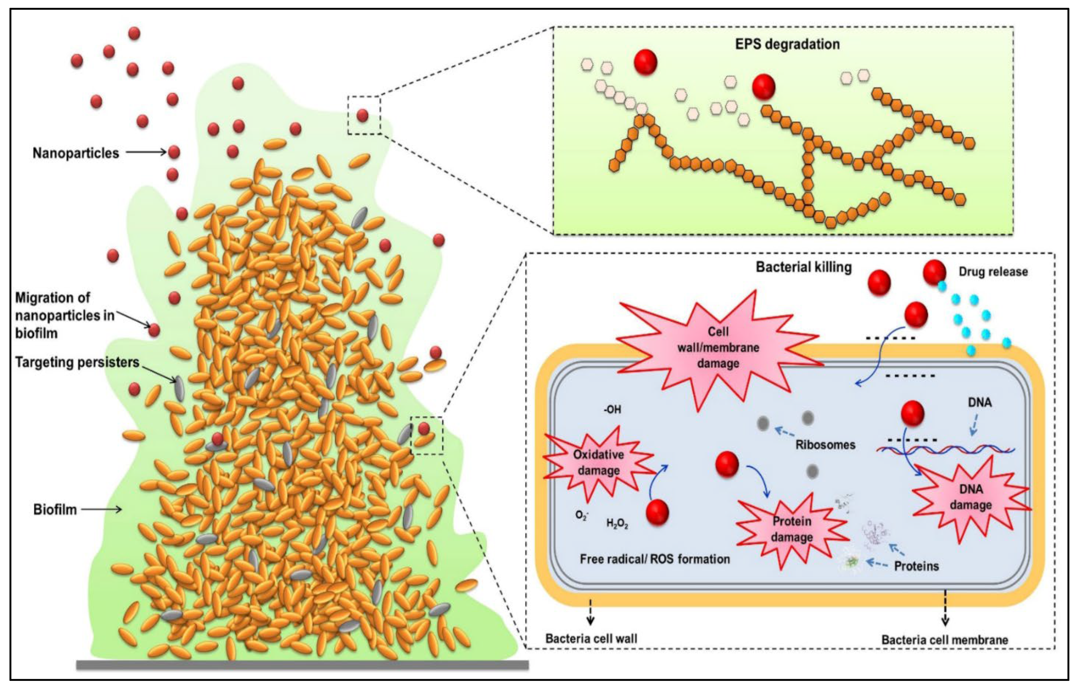
Bacteria form biofilms, which aid in adhering to surfaces and assisting in the maturation and dispersion of bacteria, both of which are required for the spread of bacteria and the creation of contamination [202]. Green synthesized Ag and ZnO NPs are the most explored nanomaterials studied for the prevention of bacterial growth and the production of biofilms. It has been reported that AgNPs synthesized by green methods using different parts of plant extracts significantly inhibit the growth of E. coli, P. aeruginosa, S. aurous, B. cereus, and K. pneumonia, and also inhibit biofilm formation [203,204,205]. In another study, it was found that biologically synthesized ZnO NPs enhanced the antimicrobial activity and inhibited the biofilm formation by E. coli and S. aureus [206,207]. L. monocytogenes is a bacterium that causes listeriosis, a foodborne disease that increases the risk of forming biofilms in the food industry and helps the bacteria to survive in challenging environments. Thus, developing antibiofilm materials for food-processing environments is highly necessary [208]. Nanomaterials with visible light illumination inactivate and reduce the biofilm of L. monocytogenes on TiO2-coated aluminum and stainless steel plates after 72-hand illumination, which is suggested as a safe and effective method for use in facilities like food processing for biofilm inhibition [208]. The combination of Au, Ag, and chitosan NPs at higher concentrations effectively inhibited biofilm growth and decreased metabolic activity (80%) in all the tested strains except for Salmonella enterica infantis; however, nanoparticles were not able to completely remove biofilm mass [209]. Biogenic AgNPs were developed from Convolvulus arvensis extract and also with chitosan to test against Gram-positive and Gram-negative bacteria. It was reported that nanoparticles decreased the bacterial cell count (S. aureus and E. coli) and also exerted antibiofilm potential against P. aeruginosa. Additionally, nanoparticles showed an anticancer effect by increasing cytotoxicity against breast cancer cell lines and altogether exerting an antibacterial, anti-biofilm, and anticancer effect [210]. A nanodrug delivery system was developed using chitosan NPs, phosphatidylcholine, and gentamicin and showed that it could effectively damage and remove the biofilm formed by pathogens via permeation through the biofilm by NPs that were engulfed by macrophages, which led to the killing of bacteria, suggesting that NPs could be a promising nano-antibacterial agent against biofilms and intracellular bacteria [211]. AgNPs were coated onto stainless steel and a polyethylene surface to test the influence of biofilm formation, which enhances the contact of microorganisms [212]. AgNP-decorated amphiphilic carbonaceous particles were developed and incorporated into urinary catheters. It improved the mechanical properties and surface wettability; possessed bacterial resistance, adhesion, and biofilm formation without inducing toxicity in rabbits; and acted as an effective antibacterial and antibiofilm agent [213]. Usnic acid-loaded chitosan NPs were prepared via simple ionic gelification and showed 24% encapsulation and 5.2% loading capacity, with a spherical and rough surface. It was found that usnic acid-chitosan NPs exerted significant antibacterial activity against persister cells and inhibited biofilm formation by Gram-negative and Gram-positive bacteria. Thus, it could be an effective alternative method for the use of usnic acid in NPs [214]. AgNPs prepared from the shoot extract of Tamarix nilotica showed that AgNPs significantly decreased the swimming, motility, cell growth, and biofilm formation of Listeria test strains. Ultimately, NPs exerted antibiofilm and wound-healing properties and could be effectively used to prevent infection with L. monocytogenes in the food industry [215]. ZnO NPs were tested against Campylobacter jejuni biofilms and it was found that NPs penetrated the biofilm within 1 h without damaging it and directly interacted with sessile cells in biofilms. NPs then induce increased free radical generation and alter DNA/RNA bases, leading to C. jejuni cell death [216]. Clove oil-loaded chitosan NPs and gelatin electrospun nanofibers were tested against biofilms of E. coli, and it was found that nanofiber treatment effectively reduced biofilm formation and maintained the color and flavor of cucumber for more than 4 days [217]. The role of nanotechnology in eradicating biofilms formed by pathogenic bacteria is shown in Figure 4.
4. Limitation, Challenges, and Future Prospectus
Nanotechnology is essential for achieving food security and can enhance crop production by controlling microbial, pest, and weed diseases with high nutritional value, security, and safety. Nanotechnology helps in the development of materials, devices, or systems at the nanometer scale and possesses a high surface-to-volume ratio with unique physiochemical properties in the agricultural sector, as reviewed above. Nanotechnology has a significant impact on the food industry because it allows for the superior processing, packaging, and long-term storage of food. This has led to an enormous increase in the food industry by improving both the flavor and texture of food. Via pathogen detection, nanomaterials and nanosensors improve security by assisting consumers and providing glimpses into the overall condition of the food inside, as well as its nutritional status. Since the majority of food bioactives that fight different diseases are hydrophobic in nature and have low bioavailability and stability, nanotechnology-based delivery methods have been able to enhance food bioactive compound bioavailability and targeted delivery [219]. Despite the many innovative products and development processes in the food industry, nanotechnology faces a significant challenge in producing edible and non-toxic nanocompounds or delivery systems that are unsafe for human consumption using cost-effective methods [220]. It has been reported that the bioaccumulation of nanomaterials, such as AgNPs derived from nanopackaging, has been observed in both food and humans [221]. As a result of their release from contaminated food or into the environment, the widespread use of nanomaterials in food packaging may be cause for concern. NPs can enter the body via ingestion, inhalation, or skin contact, and due to their nanoscale size and large surface area, they are capable of readily penetrating cells and causing toxicity in distant areas of the body [222]. At higher concentrations, NPs can cause toxicity by denaturing the body’s proteins and enzymes due to an increase in the production of free radicals, which is considered a key mechanism leading to oxidative stress in humans [223,224]. According to a recent study, inflammation, oxidative stress, genotoxicity, chromosome instability, mitochondrial damage, and apoptosis have been shown to be caused by TiO2, which is used as a food additive or in another form of food upon entering the body [225]. However, data related to the dispersion of nanoparticles from packaging materials into food and their ultimate toxicological effects are limited [226]. Moreover, the use of nanopesticides and nanofertilizers in the agriculture sector poses a serious health risk to farmers since they can contaminate water, soil, or the atmosphere. It is expected that once NPs accumulate in the soil, plant growth will be hampered, and they may accumulate in edible tissues as well [227]. As soon as the NPs are released into the agro-environment, they undergo a variety of transformations that may cause toxicity in both plants and humans [226]. Due to the inherent risk to human health posed by the use of NPs as pesticides and microbicides, strict adherence to risk assessment procedures is required when preparing food [228]. According to reports, exposure to AgNPs causes phytotoxicity in aquatic plants like Egeria densa and Juncus effusus by increasing lipid peroxidation, changing physiological traits, and causing additional stress in submerged macrophytes [229]. It has been proposed that green nanofillers are needed for nanocomposite research to ensure the safety of the environment, plants, humans, animals, and aquatic life [226]. For a safer implementation of NPs in the food sector, effective regulations, policies, and regulatory laws are required. For instance, it is important to educate the public on the effects of nanomaterials on human health, safety, and the environment. Additionally, risk analysis and safety requirements must be met before nanofood is made available in the market [230]. According to European Union regulations, any nanotechnology-based food constituent, such as nanofoods or food packaging, is required to pass a Food and Drug Administration (FDA) safety assessment before being approved for use [231]. However, many countries do not follow proper regulations for the production and processing of nanomaterials, so toxicological screening is critical for the application of nanotechnology [219]. Apart from nanotoxicity, there are practical limitations to developing nanocoating strategies to regulate the surface properties of substrates for application in the environment, biosystems, food systems, etc. The coating of fruits is time-consuming and the desired coating is not achievable. Thus, rapid and novel coating strategies are highly demanded [91]). Upon genetic editing of plants, NP-loaded cargo size and amount for CRISPR DNA and protein cargoes have been well established. Additionally, it is not clearly understood whether NPs can carry CRISPR reagents to plant mitochondria for genome modification. It remains unknown whether these NPs can carry CRISPR reagents to plant plastids and mitochondria for genome modification [30].
5. Conclusions
Recently, nanotechnology has provided a path for increasing food availability and developing new agricultural products, particularly by minimizing nutrient losses from fertilizers and improving food yield through pest and nutrient management. Nanofertilizer improved seedling vigor index, increased reserve food-mobilizing enzyme activities, increased antioxidant enzymes, decreased lipid peroxidation, stimulated chlorophyll content, and effectively enhanced crop yield by promoting source activity. Nanotechnology helps in food processing, food modification, stability, sensing, enhanced shelf life, and decreased food losses. Nanomaterials protect plants against pathogens by inhibiting microbial growth, inducing innate plant immunity, and delivering pesticides and micronutrients. Furthermore, NPs improve stress tolerance by reducing abscisic acid, regulating gibberellic acid, and improving seed germination. Nanoencapsulation helps regulate and release the active ingredients; protects them from chemicals, heat, and moisture; and enhances thermal stability and photostability. Nanotechnology-based foods challenge both the private sector and the government, assuring confidence in consumers and accepting nanofoods in the marketplace. In recent years, it has been extensively reported that nanocolloidal particles are actively utilized in various facets of the food industry, including the safety and quality of food, nutrition, processing, and packaging. The structure and function of colloidal particles are vital for the development of foods that are safer, more nutritious, of higher quality, and more sustainable. The use of NPs in food packaging has fewer health risks than their use as ingredients in food. During the process of manufacturing and application, nanomaterials pose a constant risk of entering the food chain via water, air, and soil. This can result in DNA damage, disruption of cell membranes, and cell death. Furthermore, in vivo research on the effects of nanofoods on human and animal health has been extremely limited. Therefore, it is highly recommended to conduct increasing in vivo research on the effects of nanofoods to determine their potential hazards at the molecular and genetic levels. Additionally, future investigations could also control the potential hazards posed by nanoparticles or nanocomposites via the application of sustainable synthesis techniques and searching for simple, rapid, ecofriendly, and inexpensive strategies for the breakdown and removal of existing nanomaterials at attack sites. Furthermore, many nations lack the appropriate rules and laws for the manufacture and handling of nanomaterials. Therefore, comprehensive federal laws and regulations, along with a strict toxicological assessment system, are urgently required for the legitimate utilization of nanotechnology in the food manufacturing industry. Thus, the use of the aforementioned nanotechnologies could significantly enhance the production and processing of foods and the quality of products, which will improve human health and well-being if handled and governed effectively.
Supplementary Materials
The following supporting information can be downloaded at: https://www.mdpi.com/article/10.3390/plants12132565/s1, Supplementary Table S1: Nanoparticles employed for the detection of foodborne pathogens; Supplementary Table S2: Nanomaterial-based biosensors applied in food science and food nanotechnology. References [232,233,234,235,236,237,238,239,240,241,242,243,244,245,246,247,248,249,250,251,252,253,254,255,256,257,258,259,260,261,262,263,264,265,266,267,268,269,270,271,272,273,274] are also cited in Supplementary Materials.
Funding
This research received no external funding.
Data Availability Statement
Not applicable.
Conflicts of Interest
The authors declare no conflict of interest.
References
- Fiaz, S.; Noor, M.A.; Aldosri, F.O. Achieving food security in the Kingdom of Saudi Arabia through innovation: Potential role of agricultural extension. J. Saudi Soc. Agric. Sci. 2018, 17, 365–375. [Google Scholar] [CrossRef] [Green Version]
- Alsubaie, A.S.R.; Berekaa, M.M. Food Safety in Saudi Arabia: A Public Health Priority. Ann. Med. Health Sci. Res. 2020, 10, 1142–1147. [Google Scholar]
- Jones, K.E.; Patel, N.G.; Levy, M.A.; Storeygard, A.; Balk, D.; Gittleman, J.L.; Daszak, P. Global trends in emerging infectious diseases. Nature 2008, 451, 990–993. [Google Scholar] [CrossRef] [PubMed]
- Handford, C.E.; Dean, M.; Henchion, M.; Spence, M.; Elliott, C.T.; Campbell, K. Implications of nanotechnology for the agri-food industry: Opportunities, benefits and risks. Trends Food Sci. Technol. 2014, 40, 226–241. [Google Scholar] [CrossRef]
- Bata-Vidács, I.; Adányi, N.; Beczner, J.; Farkas, J.; Székács, A. Nanotechnology and microbial food safety. In Microbial Pathogens and Strategies for Combating Them: Science, Technology and Education; Méndez-Vilas, A., Ed.; Formatex Research Center: Badajoz, Spain, 2013; pp. 155–159. [Google Scholar]
- Ansari, M.A.; Akhtar, S.; Rauf, M.A.; Alomary, M.N.; AlYahya, S.; Alghamdi, S.; Almessiere, M.A.; Baykal, A.; Khan, F.; Adil, S.F.; et al. Sol–Gel Synthesis of Dy-Substituted Ni0.4Cu0.2Zn0.4 (Fe2-xDyx) O4 Nano Spinel Ferrites and Evaluation of Their Antibacterial, Antifungal, Antibiofilm and Anticancer Potentialities for Biomedical Application. Int. J. Nanomed. 2021, 16, 5633. [Google Scholar] [CrossRef]
- Srinivasa, C.; Kumar, S.S.; Pradeep, S.; Prasad, S.K.; Veerapur, R.; Ansari, M.A.; Alomary, M.N.; Alghamdi, S.; Almehmadi, M.; Gc, K.; et al. Eco-Friendly Synthesis of MnO2 Nanorods Using Gmelina arborea Fruit Extract and Its Anticancer Potency Against MCF-7 Breast Cancer Cell Line. Int. J. Nanomed. 2022, 17, 901–907. [Google Scholar] [CrossRef]
- Jagtap, R.R.; Garud, A.; Puranik, S.S.; Rudrapal, M.; Ansari, M.A.; Alomary, M.N.; Alshamrani, M.; Salawi, A.; Almoshari, Y.; Khan, J.; et al. Biofabrication of Silver Nanoparticles (AgNPs) Using Embelin for Effective Therapeutic Management of Lung Cancer. Front. Nutr. 2022, 9, 960674. [Google Scholar] [CrossRef]
- Murali, M.; Kalegowda, N.; Gowtham, H.G.; Ansari, M.A.; Alomary, M.N.; Alghamdi, S.; Shilpa, N.; Singh, S.B.; Thriveni, M.C.; Aiyaz, M.; et al. Plant-Mediated Zinc Oxide NPs: Advances in the New Millennium towards Understanding Their Therapeutic Role in Biomedical Applications. Pharmaceutics 2021, 13, 1662. [Google Scholar] [CrossRef]
- Govindasamy, R.; Govindarasu, M.; Alharthi, S.S.; Mani, P.; Bernaurdshaw, N.; Gomathi, T.; Ansari, M.A.; Alomary, M.N.; Atwah, B.; Malik, M.S.; et al. Sustainable green synthesis of yttrium oxide (Y2O3) NPs using Lantana camara leaf extracts: Physicochemical characterization, photocatalytic degradation, antibacterial, and anticancer potency. Nanomaterials 2022, 12, 2393. [Google Scholar] [CrossRef]
- Ray, S.S.; Bandyopadhyay, J. Nanotechnology-enabled biomedical engineering: Current trends, future scopes, and perspectives. Nanotechnol. Rev. 2021, 10, 728–743. [Google Scholar] [CrossRef]
- Mazayen, Z.M.; Ghoneim, A.M.; Elbatanony, R.S.; Basalious, E.B.; Bendas, E.R. Pharmaceutical nanotechnology: From the bench to the market. Futur. J. Pharm. Sci. 2022, 8, 1–11. [Google Scholar] [CrossRef]
- Farooq, M.A.; Hannan, F.; Islam, F.; Ayyaz, A.; Zhang, N.; Chen, W.; Zhang, K.; Huang, Q.; Xu, L.; Zhou, W. The potential of nanomaterials for sustainable modern agriculture: Present findings and future perspectives. Environ. Sci. Nano 2022, 9, 1926–1951. [Google Scholar] [CrossRef]
- Gupta, A.; Eral, H.B.; Hatton, T.A.; Doyle, P.S. Nanoemulsions: Formation, properties and applications. Soft Matter 2016, 12, 2826–2841. [Google Scholar] [CrossRef] [PubMed] [Green Version]
- Chaudhry, Q.; Scotter, M.; Blackburn, J.; Ross, B.; Boxall, A.; Castle, L.; Aitken, R.; Watkins, R. Applications and implications of nanotechnologies for the food sector. Food Addit. Contam. Part A 2008, 25, 241–258. [Google Scholar] [CrossRef] [PubMed]
- Summit, W.F. Rome Declaration on World Food Security and World Food Summit Plan of Action; FAO: Rome, Italy, 1996. [Google Scholar]
- Schmidhuber, J.; Tubiello, F.N. Global food security under climate change. Proc. Natl. Acad. Sci. USA 2007, 104, 19703–19708. [Google Scholar] [CrossRef] [PubMed]
- Duro, J.A.; Lauk, C.; Kastner, T.; Erb, K.-H.; Haberl, H. Global inequalities in food consumption, cropland demand and land-use efficiency: A decomposition analysis. Glob. Environ. Chang. 2020, 64, 102124. [Google Scholar] [CrossRef]
- UN News. UN Report: One-Third of World’s Food Wasted Annually, at Great Economic, Environmental Cost. 2013. Available online: http://www.un.org/apps/news/story.asp?NewsID=45816#.VtR44niOeLE (accessed on 5 June 2023).
- Food Wastage Footprint. Impacts on Natural Resources. Summary Report. 2013. Available online: http://www.fao.org/docrep/018/i3347e/i3347e.pdf (accessed on 5 June 2023).
- Shukla, P.S.; Mantin, E.G.; Adil, M.; Bajpai, S.; Critchley, A.T.; Prithiviraj, B. Ascophyllum nodosum-Based Biostimulants: Sustainable Applications in Agriculture for the Stimulation of Plant Growth, Stress Tolerance, and Disease Management. Front. Plant Sci. 2019, 10, 655. [Google Scholar] [CrossRef] [Green Version]
- Dos Reis, S.P.; Lima, A.M.; de Souza, C.R.B. Recent Molecular Advances on Downstream Plant Responses to Abiotic Stress. Int. J. Mol. Sci. 2012, 13, 8628–8647. [Google Scholar] [CrossRef]
- Kagan, C.R. At the Nexus of Food Security and Safety: Opportunities for Nanoscience and Nanotechnology. ACS Nano 2016, 10, 2985–2986. [Google Scholar] [CrossRef] [Green Version]
- Pal, M. Nanotechnology: A New Approach in Food Packaging. J. Food Microbiol. Saf. Hyg. 2017, 2, 1–2. [Google Scholar] [CrossRef]
- Wesley, S.J.; Raja, P.; Raj, A.; Tiroutchelvamae, D. Review on—Nanotechnology Applications in Food Packaging and Safety. Int. J. Eng. Res. 2014, 3, 645–651. [Google Scholar] [CrossRef]
- Alrobaish, W.S.; Vlerick, P.; Luning, P.A.; Jacxsens, L. Food safety governance in Saudi Arabia: Challenges in control of imported food. J. Food Sci. 2020, 86, 16–30. [Google Scholar] [CrossRef] [PubMed]
- Inbaraj, B.S.; Chen, B. Nanomaterial-based sensors for detection of foodborne bacterial pathogens and toxins as well as pork adulteration in meat products. J. Food Drug Anal. 2015, 24, 15–28. [Google Scholar] [CrossRef]
- Sadeghi, R.; Rodriguez, R.J.; Yao, Y.; Kokini, J.L. Advances in Nanotechnology as They Pertain to Food and Agriculture: Benefits and Risks. Annu. Rev. Food Sci. Technol. 2017, 8, 467–492. [Google Scholar] [CrossRef] [PubMed]
- Rizvi, S.S.; Moraru, C.I.; Bouwmeester, H.; Kampers, F.W.; Cheng, Y. Nanotechnology and food safety. In Ensuring Global Food Safety; Academic Press: Cambridge, MA, USA, 2022; pp. 325–340. [Google Scholar]
- Demirer, G.S.; Silva, T.N.; Jackson, C.T.; Thomas, J.B.; Ehrhardt, D.W.; Rhee, S.Y.; Mortimer, J.C.; Landry, M.P. Nanotechnology to advance CRISPR–Cas genetic engineering of plants. Nat. Nanotechnol. 2021, 16, 243–250. [Google Scholar] [CrossRef] [PubMed]
- Kumar, S.; Nehra, M.; Dilbaghi, N.; Marrazza, G.; Hassan, A.A.; Kim, K.-H. Nano-based smart pesticide formulations: Emerging opportunities for agriculture. J. Control. Release 2019, 294, 131–153. [Google Scholar] [CrossRef]
- Sharma, G.; Kumar, A.; Devi, K.A.; Prajapati, D.; Bhagat, D.; Pal, A.; Raliya, R.; Biswas, P.; Saharan, V. Chitosan nanofertilizer to foster source activity in maize. Int. J. Biol. Macromol. 2020, 145, 226–234. [Google Scholar] [CrossRef]
- El-Naggar, M.E.; Abdelsalam, N.R.; Fouda, M.M.; Mackled, M.I.; Al-Jaddadi, M.A.; Ali, H.M.; Siddiqui, M.H.; Kandil, E.E. Soil Application of Nano Silica on Maize Yield and Its Insecticidal Activity against Some Stored Insects after the Post-Harvest. Nanomaterials 2020, 10, 739. [Google Scholar] [CrossRef] [Green Version]
- Abdel-Aziz, H.M.M.; Soliman, M.I.; Al-Saoud, A.M.A.; El-Sherbeny, G.A. Waste-Derived NPK Nanofertilizer Enhances Growth and Productivity of Capsicum annuum L. Plants 2021, 10, 1144. [Google Scholar] [CrossRef]
- Mosa, W.F.A.; El-Shehawi, A.M.; Mackled, M.I.; Salem, M.Z.M.; Ghareeb, R.Y.; Hafez, E.E.; Behiry, S.I.; Abdelsalam, N.R. Productivity performance of peach trees, insecticidal and antibacterial bioactivities of leaf extracts as affected by nanofertilizers foliar application. Sci. Rep. 2021, 11, 10205. [Google Scholar] [CrossRef]
- Atteya, A.K.G.; Albalawi, A.N.; El-Serafy, R.S.; Albalawi, K.N.; Bayomy, H.M.; Genaidy, E.A.E. Response of Moringa oleifera Seeds and Fixed Oil Production to Vermicompost and NPK Fertilizers under Calcareous Soil Conditions. Plants 2021, 10, 1998. [Google Scholar] [CrossRef]
- Al-Amri, N.; Tombuloglu, H.; Slimani, Y.; Akhtar, S.; Barghouthi, M.; Almessiere, M.; Alshammari, T.; Baykal, A.; Sabit, H.; Ercan, I.; et al. Size effect of iron (III) oxide nanomaterials on the growth, and their uptake and translocation in common wheat (Triticum aestivum L.). Ecotoxicol. Environ. Saf. 2020, 194, 110377. [Google Scholar] [CrossRef] [PubMed]
- Tombuloglu, H.; Albenayyan, N.; Slimani, Y.; Akhtar, S.; Tombuloglu, G.; Almessiere, M.; Baykal, A.; Ercan, I.; Sabit, H.; Manikandan, A. Fate and impact of maghemite (γ-Fe2O3) and magnetite (Fe3O4) nanoparticles in barley (Hordeum vulgare L.). Environ. Sci. Pollut. Res. 2021, 29, 4710–4721. [Google Scholar] [CrossRef] [PubMed]
- Abdelmigid, H.M.; Morsi, M.M.; Hussien, N.A.; Alyamani, A.A.; Alhuthal, N.A.; Albukhaty, S. Green Synthesis of Phosphorous-Containing Hydroxyapatite NPs (nHAP) as a Novel Nano-Fertilizer: Preliminary Assessment on Pomegranate (Punica granatum L.). Nanomaterials 2022, 12, 1527. [Google Scholar] [CrossRef]
- El-Gioushy, S.F.; Ding, Z.; Bahloul, A.M.E.; Gawish, M.S.; Abou El Ghit, H.M.; Abdelaziz, A.M.R.A.; El-Desouky, H.S.; Sami, R.; Khojah, E.; Hashim, T.A.; et al. Foliar Application of Nano, Chelated, and Conventional Iron Forms Enhanced Growth, Nutritional Status, Fruiting Aspects, and Fruit Quality of Washington Navel Orange Trees (Citrus sinensis L. Osbeck). Plants 2021, 10, 2577. [Google Scholar] [CrossRef] [PubMed]
- Ahmed, T.; Wu, Z.; Jiang, H.; Luo, J.; Noman, M.; Shahid, M.; Manzoor, I.; Allemailem, K.S.; Alrumaihi, F.; Li, B. Bioinspired Green Synthesis of Zinc Oxide NPs from a Native Bacillus cereus Strain RNT6: Characterization and Antibacterial Activity against Rice Panicle Blight Pathogens Burkholderia glumae and B. gladioli. Nanomaterials 2021, 11, 884. [Google Scholar] [CrossRef] [PubMed]
- Jiang, H.; Lv, L.; Ahmed, T.; Jin, S.; Shahid, M.; Noman, M.; Osman, H.-E.H.; Wang, Y.; Sun, G.; Li, X.; et al. Effect of the Nanoparticle Exposures on the Tomato Bacterial Wilt Disease Control by Modulating the Rhizosphere Bacterial Community. Int. J. Mol. Sci. 2021, 23, 414. [Google Scholar] [CrossRef]
- Paulraj, M.G.; Ignacimuthu, S.; Gandhi, M.R.; Shajahan, A.; Ganesan, P.; Packiam, S.M.; Al-Dhabi, N.A. Comparative studies of tripolyphosphate and glutaraldehyde cross-linked chitosan-botanical pesticide nanoparticles and their agricultural applications. Int. J. Biol. Macromol. 2017, 104, 1813–1819. [Google Scholar] [CrossRef]
- Sujitha, V.; Murugan, K.; Dinesh, D.; Pandiyan, A.; Aruliah, R.; Hwang, J.-S.; Kalimuthu, K.; Panneerselvam, C.; Higuchi, A.; Aziz, A.T.; et al. Green-synthesized CdS nano-pesticides: Toxicity on young instars of malaria vectors and impact on enzymatic activities of the non-target mud crab Scylla serrata. Aquat. Toxicol. 2017, 188, 100–108. [Google Scholar] [CrossRef]
- Hofmann, T.; Lowry, G.V.; Ghoshal, S.; Tufenkji, N.; Brambilla, D.; Dutcher, J.R.; Gilbertson, L.M.; Giraldo, J.P.; Kinsella, J.M.; Landry, M.P.; et al. Technology readiness and overcoming barriers to sustainably implement nanotechnology-enabled plant agriculture. Nat. Food 2020, 1, 416–425. [Google Scholar] [CrossRef]
- Doyle, C.; Higginbottom, K.; Swift, T.A.; Winfield, M.; Bellas, C.; Benito-Alifonso, D.; Fletcher, T.; Galan, M.C.; Edwards, K.; Whitney, H.M. A simple method for spray-on gene editing in planta. bioRxiv 2019, 805036. [Google Scholar] [CrossRef] [Green Version]
- Yong, J.; Zhang, R.; Bi, S.; Li, P.; Sun, L.; Mitter, N.; Carroll, B.J.; Xu, Z.P. Sheet-like clay nanoparticles deliver RNA into developing pollen to efficiently silence a target gene. Plant Physiol. 2021, 187, 886–899. [Google Scholar] [CrossRef] [PubMed]
- Kwak, S.-Y.; Lew, T.T.S.; Sweeney, C.J.; Koman, V.B.; Wong, M.H.; Bohmert-Tatarev, K.; Snell, K.D.; Seo, J.S.; Chua, N.-H.; Strano, M.S. Chloroplast-selective gene delivery and expression in planta using chitosan-complexed single-walled carbon nanotube carriers. Nat. Nanotechnol. 2019, 14, 447–455. [Google Scholar] [CrossRef]
- Fu, L.; Wang, Z.; Dhankher, O.P.; Xing, B. Nanotechnology as a new sustainable approach for controlling crop diseases and increasing agricultural production. J. Exp. Bot. 2020, 71, 507–519. [Google Scholar] [CrossRef] [PubMed]
- Alghuthaymi, M.A.; Kalia, A.; Bhardwaj, K.; Bhardwaj, P.; Abd-Elsalam, K.A.; Valis, M.; Kuca, K. Nanohybrid Antifungals for Control of Plant Diseases: Current Status and Future Perspectives. J. Fungi 2021, 7, 48. [Google Scholar] [CrossRef]
- Pérez-De-Luque, A.; Rubiales, D. Nanotechnology for parasitic plant control. Pest Manag. Sci. 2009, 65, 540–545. [Google Scholar] [CrossRef]
- Li, Y.; Zhang, P.; Li, M.; Shakoor, N.; Adeel, M.; Zhou, P.; Guo, M.; Jiang, Y.; Zhao, W.; Lou, B.; et al. Application and mechanisms of metal-based nanoparticles in the control of bacterial and fungal crop diseases. Pest Manag. Sci. 2023, 79, 21–36. [Google Scholar] [CrossRef]
- Khan, S.T.; Ahmad, J.; Ahamed, M.; Jousset, A. Sub-lethal doses of widespread nanoparticles promote antifungal activity in Pseudomonas protegens CHA0. Sci. Total. Environ. 2018, 627, 658–662. [Google Scholar] [CrossRef]
- Abdullahi, A.; Ahmad, K.; Ismail, I.S.; Asib, N.; Ahmed, O.H.; Abubakar, A.I.; Siddiqui, Y.; Ismail, M.R. Potential of Using Ginger Essential Oils-Based Nanotechnology to Control Tropical Plant Diseases. Plant Pathol. J. 2020, 36, 515–535. [Google Scholar] [CrossRef]
- Mitter, N.; Worrall, E.A.; Robinson, K.E.; Li, P.; Jain, R.G.; Taochy, C.; Fletcher, S.J.; Carroll, B.J.; Lu, G.Q.; Xu, Z.P. Clay nanosheets for topical delivery of RNAi for sustained protection against plant viruses. Nat. Plants 2017, 3, 16207. [Google Scholar] [CrossRef]
- Tauseef, A.; Hisamuddin; Khalilullah, A.; Uddin, I. Role of MgO nanoparticles in the suppression of Meloidogyne incognita, infecting cowpea and improvement in plant growth and physiology. Exp. Parasitol. 2020, 220, 108045. [Google Scholar] [CrossRef] [PubMed]
- Ntasiou, P.; Kerou, A.K.; Karamanidou, T.; Vlachou, A.; Tziros, G.T.; Tsouknidas, A.; Karaoglanidis, G.S. Synthesis and Characterization of Novel Copper Nanoparticles for the Control of Leaf Spot and Anthracnose Diseases of Olive. Nanomaterials 2021, 11, 1667. [Google Scholar] [CrossRef] [PubMed]
- Vanti, G.L.; Masaphy, S.; Kurjogi, M.; Chakrasali, S.; Nargund, V.B. Synthesis and application of chitosan-copper nanoparticles on damping off causing plant pathogenic fungi. Int. J. Biol. Macromol. 2019, 156, 1387–1395. [Google Scholar] [CrossRef]
- El-Abeid, S.E.; Ahmed, Y.; Daròs, J.-A.; Mohamed, M.A. Reduced Graphene Oxide Nanosheet-Decorated Copper Oxide Nanoparticles: A Potent Antifungal Nanocomposite against Fusarium Root Rot and Wilt Diseases of Tomato and Pepper Plants. Nanomaterials 2020, 10, 1001. [Google Scholar] [CrossRef] [PubMed]
- Guo, Y.; Khan, R.A.A.; Xiong, Y.; Fan, Z. Enhanced suppression of soil-borne phytopathogenic bacteria Ralstonia solanacearum in soil and promotion of tomato plant growth by synergetic effect of green synthesized nanoparticles and plant extract. J. Appl. Microbiol. 2022, 132, 3694–3704. [Google Scholar] [CrossRef]
- Zudyte, B.; Luksiene, Z. Visible light-activated ZnO nanoparticles for microbial control of wheat crop. J. Photochem. Photobiol. B Biol. 2021, 219, 112206. [Google Scholar] [CrossRef]
- Dawoud, T.M.; Yassin, M.A.; El-Samawaty, A.R.M.; Elgorban, A.M. Silver nanoparticles synthesized by Nigrospora oryzae showed antifungal activity. Saudi J. Biol. Sci. 2020, 28, 1847–1852. [Google Scholar] [CrossRef]
- Sofy, A.R.; Sofy, M.R.; Hmed, A.A.; Dawoud, R.A.; Alnaggar, A.E.-A.M.; Soliman, A.M.; El-Dougdoug, N.K. Ameliorating the Adverse Effects of Tomato mosaic tobamovirus Infecting Tomato Plants in Egypt by Boosting Immunity in Tomato Plants Using Zinc Oxide Nanoparticles. Molecules 2021, 26, 1337. [Google Scholar] [CrossRef]
- Kabeerdass, N.; Al Otaibi, A.; Rajendran, M.; Manikandan, A.; Kashmery, H.A.; Rahman, M.M.; Madhu, P.; Khan, A.; Asiri, A.M.; Mathanmohun, M. Bacillus-Mediated Silver Nanoparticle Synthesis and Its Antagonistic Activity against Bacterial and Fungal Pathogens. Antibiotics 2021, 10, 1334. [Google Scholar] [CrossRef]
- Mostafa, Y.S.; Alamri, S.A.; Alrumman, S.A.; Hashem, M.; Baka, Z.A. Green Synthesis of Silver Nanoparticles Using Pomegranate and Orange Peel Extracts and Their Antifungal Activity against Alternaria solani, the Causal Agent of Early Blight Disease of Tomato. Plants 2021, 10, 2363. [Google Scholar] [CrossRef]
- Iliger, K.S.; Sofi, T.A.; Bhat, N.A.; Ahanger, F.A.; Sekhar, J.C.; Elhendi, A.Z.; Al-Huqail, A.A.; Khan, F. Copper nanoparticles: Green synthesis and managing fruit rot disease of chilli caused by Colletotrichum capsici. Saudi J. Biol. Sci. 2021, 28, 1477–1486. [Google Scholar] [CrossRef] [PubMed]
- Abdelaziz, A.M.; Salem, S.S.; Khalil, A.M.A.; El-Wakil, D.A.; Fouda, H.M.; Hashem, A.H. Potential of biosynthesized zinc oxide NPs to control Fusarium wilt disease in eggplant (Solanum melongena) and promote plant growth. Biometals 2022, 35, 601–616. [Google Scholar] [CrossRef]
- Zaki, S.A.; Ouf, S.A.; Albarakaty, F.M.; Habeb, M.M.; Aly, A.A.; Abd-Elsalam, K.A. Trichoderma harzianum-mediated ZnO NPs: A green tool for controlling soil-borne pathogens in cotton. J. Fungi 2021, 7, 952. [Google Scholar] [CrossRef]
- Abdelkhalek, A.; Qari, S.H.; Abu-Saied, M.A.A.-R.; Khalil, A.M.; Younes, H.A.; Nehela, Y.; Behiry, S.I. Chitosan Nanoparticles Inactivate Alfalfa Mosaic Virus Replication and Boost Innate Immunity in Nicotiana glutinosa Plants. Plants 2021, 10, 2701. [Google Scholar] [CrossRef]
- Abdelrhim, A.S.; Mazrou, Y.S.A.; Nehela, Y.; Atallah, O.O.; El-Ashmony, R.M.; Dawood, M.F.A. Silicon Dioxide Nanoparticles Induce Innate Immune Responses and Activate Antioxidant Machinery in Wheat Against Rhizoctonia solani. Plants 2021, 10, 2758. [Google Scholar] [CrossRef]
- Hashem, A.H.; Abdelaziz, A.M.; Askar, A.A.; Fouda, H.M.; Khalil, A.M.A.; Abd-Elsalam, K.A.; Khaleil, M.M. Bacillus megaterium-Mediated Synthesis of Selenium Nanoparticles and Their Antifungal Activity against Rhizoctonia solani in Faba Bean Plants. J. Fungi 2021, 7, 195. [Google Scholar] [CrossRef]
- Ali, S.G.; Ansari, M.A.; Alzohairy, M.A.; Alomary, M.N.; AlYahya, S.; Jalal, M.; Khan, H.M.; Asiri, S.M.M.; Ahmad, W.; Mahdi, A.A.; et al. Biogenic Gold Nanoparticles as Potent Antibacterial and Antibiofilm Nano-Antibiotics against Pseudomonas aeruginosa. Antibiotics 2020, 9, 100. [Google Scholar] [CrossRef] [PubMed] [Green Version]
- El-Badri, A.M.; Batool, M.; Wang, C.; Hashem, A.M.; Tabl, K.M.; Nishawy, E.; Kuai, J.; Zhou, G.; Wang, B. Selenium and zinc oxide nanoparticles modulate the molecular and morpho-physiological processes during seed germination of Brassica napus under salt stress. Ecotoxicol. Environ. Saf. 2021, 225, 112695. [Google Scholar] [CrossRef] [PubMed]
- Gohari, G.; Panahirad, S.; Sepehri, N.; Akbari, A.; Zahedi, S.M.; Jafari, H.; Dadpour, M.R.; Fotopoulos, V. Enhanced tolerance to salinity stress in grapevine plants through application of carbon quantum dots functionalized by proline. Environ. Sci. Pollut. Res. 2021, 28, 42877–42890. [Google Scholar] [CrossRef]
- Palmqvist, N.M.; Seisenbaeva, G.A.; Svedlindh, P.; Kessler, V.G. Maghemite Nanoparticles Acts as Nanozymes, Improving Growth and Abiotic Stress Tolerance in Brassica napus. Nanoscale Res. Lett. 2017, 12, 631. [Google Scholar] [CrossRef] [Green Version]
- El-Gazzar, N.; Almaary, K.; Ismail, A.; Polizzi, G. Influence of Funneliformis mosseae enhanced with titanium dioxide nanoparticles (TiO2NPs) on Phaseolus vulgaris L. under salinity stress. PLoS ONE 2020, 15, e0235355. [Google Scholar] [CrossRef] [PubMed]
- Sheikhalipour, M.; Esmaielpour, B.; Behnamian, M.; Gohari, G.; Giglou, M.T.; Vachova, P.; Rastogi, A.; Brestic, M.; Skalicky, M. Chitosan–Selenium Nanoparticle (Cs–Se NP) Foliar Spray Alleviates Salt Stress in Bitter Melon. Nanomaterials 2021, 11, 684. [Google Scholar] [CrossRef] [PubMed]
- Wang, A.; Li, J.; Al-Huqail, A.A.; Al-Harbi, M.S.; Ali, E.F.; Wang, J.; Ding, Z.; Rekaby, S.A.; Ghoneim, A.M.; Eissa, M.A. Mechanisms of Chitosan NPs in the Regulation of Cold Stress Resistance in Banana Plants. Nanomaterials 2021, 11, 2670. [Google Scholar] [CrossRef]
- Banerjee, A.; Roychoudhury, A. Maghemite nano-fertilization promotes fluoride tolerance in rice by restoring grain yield and modulating the ionome and physiome. Ecotoxicol. Environ. Saf. 2021, 215, 112055. [Google Scholar] [CrossRef]
- El-Saber, M.M.; Mahdi, A.A.; Hassan, A.H.; Farroh, K.Y.; Osman, A. Effects of magnetite NPs on physiological processes to alleviate salinity induced oxidative damage in wheat. J. Sci. Food Agric. 2021, 101, 5550–5562. [Google Scholar] [CrossRef] [PubMed]
- Wahid, I.; Rani, P.; Kumari, S.; Ahmad, R.; Hussain, S.J.; Alamri, S.; Tripathy, N.; Khan, M.I.R. Biosynthesized gold nanoparticles maintained nitrogen metabolism, nitric oxide synthesis, ions balance, and stabilizes the defense systems to improve salt stress tolerance in wheat. Chemosphere 2021, 287, 132142. [Google Scholar] [CrossRef]
- Rossi, L.; Zhang, W.; Ma, X. Cerium oxide nanoparticles alter the salt stress tolerance of Brassica napus L. by modifying the formation of root apoplastic barriers. Environ. Pollut. 2017, 229, 132–138. [Google Scholar] [CrossRef]
- Feng, Y.; Zhong, L.; Bilal, M.; Tan, Z.; Hou, Y.; Jia, S.; Cui, J. Enzymes@ZIF-8 Nanocomposites with Protection Nanocoating: Stability and Acid-Resistant Evaluation. Polymers 2018, 11, 27. [Google Scholar] [CrossRef] [Green Version]
- Yang, K.; Wang, Q.; Wang, Y.; Li, S.; Gu, Y.; Gao, N.; Zhang, F.; Lei, P.; Wang, R.; Xu, H. Poly(γ-glutamic acid) Nanocoating To Enhance the Viability of Pseudomonas stutzeri NRCB010 through Cell Surface Engineering. ACS Appl. Mater. Interfaces 2021, 13, 39957–39966. [Google Scholar] [CrossRef]
- Yemmireddy, V.K.; Farrell, G.D.; Hung, Y.-C. Development of Titanium Dioxide (TiO2) Nanocoatings on Food Contact Surfaces and Method to Evaluate Their Durability and Photocatalytic Bactericidal Property. J. Food Sci. 2015, 80, N1903–N1911. [Google Scholar] [CrossRef]
- Hannon, J.C.; Kerry, J.P.; Cruz-Romero, M.; Azlin-Hasim, S.; Morris, M.; Cummins, E. Human exposure assessment of silver and copper migrating from an antimicrobial nanocoated packaging material into an acidic food simulant. Food Chem. Toxicol. 2016, 95, 128–136. [Google Scholar] [CrossRef]
- Hu, X.; Saravanakumar, K.; Sathiyaseelan, A.; Wang, M.-H. Chitosan nanoparticles as edible surface coating agent to preserve the fresh-cut bell pepper (Capsicum annuum L. var. grossum (L.) Sendt). Int. J. Biol. Macromol. 2020, 165, 948–957. [Google Scholar] [CrossRef] [PubMed]
- Azlin-Hasim, S.; Cruz-Romero, M.C.; A Morris, M.; Cummins, E.; Kerry, J.P. Spray coating application for the development of nanocoated antimicrobial low-density polyethylene films to increase the shelf life of chicken breast fillets. Food Sci. Technol. Int. 2018, 24, 688–698. [Google Scholar] [CrossRef] [PubMed]
- Li, Y.; Rokayya, S.; Jia, F.; Nie, X.; Xu, J.; Elhakem, A.; Almatrafi, M.; Benajiba, N.; Helal, M. Shelf-life, quality, safety evaluations of blueberry fruits coated with chitosan nano-material films. Sci. Rep. 2021, 11, 55. [Google Scholar] [CrossRef]
- Correa-Pacheco, Z.N.; Corona-Rangel, M.L.; Bautista-Baños, S.; Ventura-Aguilar, R.I. Application of natural-based nanocoatings for extending the shelf life of green bell pepper fruit. J. Food Sci. 2020, 86, 95–102. [Google Scholar] [CrossRef]
- Park, J.H.; Choi, S.; Moon, H.C.; Seo, H.; Kim, J.Y.; Hong, S.P.; Lee, B.S.; Kang, E.; Lee, J.; Ryu, D.H.; et al. Antimicrobial spray nanocoating of supramolecular Fe(III)-tannic acid metal-organic coordination complex: Applications to shoe insoles and fruits. Sci. Rep. 2017, 7, 6980. [Google Scholar] [CrossRef] [PubMed] [Green Version]
- Hernández-López, G.; Ventura-Aguilar, R.I.; Correa-Pacheco, Z.N.; Bautista-Baños, S.; Barrera-Necha, L.L. Nanostructured chitosan edible coating loaded with α-pinene for the preservation of the postharvest quality of Capsicum annuum L. and Alternaria alternata control. Int. J. Biol. Macromol. 2020, 165, 1881–1888. [Google Scholar] [CrossRef]
- Karimirad, R.; Behnamian, M.; Dezhsetan, S.; Sonnenberg, A. Chitosan nanoparticles-loaded Citrus aurantium essential oil: A novel delivery system for preserving the postharvest quality of Agaricus bisporus. J. Sci. Food Agric. 2018, 98, 5112–5119. [Google Scholar] [CrossRef]
- Hossain, A.S.; Uddin, M.M.; Veettil, V.N.; Fawzi, M. Nano-cellulose based nano-coating biomaterial dataset using corn leaf biomass: An innovative biodegradable plant biomaterial. Data Brief 2018, 17, 162–168. [Google Scholar] [CrossRef]
- Altpeter, F.; Springer, N.M.; Bartley, L.E.; Blechl, A.E.; Brutnell, T.P.; Citovsky, V.; Conrad, L.J.; Gelvin, S.B.; Jackson, D.P.; Kausch, A.P.; et al. Advancing Crop Transformation in the Era of Genome Editing. Plant Cell 2016, 28, 1510–1520. [Google Scholar] [CrossRef] [Green Version]
- Mahfouz, M.M.; Piatek, A.; Stewart, C.N., Jr. Genome engineering via TALENs and CRISPR/Cas9 systems: Challenges and perspectives. Plant Biotechnol. J. 2014, 12, 1006–1014. [Google Scholar] [CrossRef]
- Martin-Ortigosa, S.; Peterson, D.J.; Valenstein, J.S.; Lin, V.S.-Y.; Trewyn, B.G.; Lyznik, L.A.; Wang, K. Mesoporous Silica Nanoparticle-Mediated Intracellular Cre Protein Delivery for Maize Genome Editing via loxP Site Excision. Plant Physiol. 2014, 164, 537–547. [Google Scholar] [CrossRef] [Green Version]
- Demirer, G.S.; Zhang, H.; Matos, J.L.; Goh, N.S.; Cunningham, F.J.; Sung, Y.; Chang, R.; Aditham, A.J.; Chio, L.; Cho, M.-J.; et al. High aspect ratio nanomaterials enable delivery of functional genetic material without DNA integration in mature plants. Nat. Nanotechnol. 2019, 14, 456–464. [Google Scholar] [CrossRef]
- Wong, M.H.; Misra, R.P.; Giraldo, J.P.; Kwak, S.Y.; Son, Y.; Landry, M.P.; Swan, J.W.; Blankschtein, D.; Strano, M.S. Lipid exchange envelope penetration (LEEP) of nanoparticles for plant engineering: A universal localization mechanism. Nano Lett. 2016, 16, 1161–1172. [Google Scholar] [CrossRef] [Green Version]
- Demirer, G.S.; Zhang, H.; Goh, N.S.; Pinals, R.L.; Chang, R.; Landry, M.P. Carbon nanocarriers deliver siRNA to intact plant cells for efficient gene knockdown. Sci. Adv. 2020, 6, eaaz0495. [Google Scholar] [CrossRef]
- Santana, I.; Wu, H.; Hu, P.; Giraldo, J.P. Targeted delivery of nanomaterials with chemical cargoes in plants enabled by a biorecognition motif. Nat. Commun. 2020, 11, 2045. [Google Scholar] [CrossRef] [PubMed]
- Schwartz, S.H.; Hendrix, B.; Hoffer, P.; Sanders, R.A.; Zheng, W. Carbon Dots for Efficient Small Interfering RNA Delivery and Gene Silencing in Plants. Plant Physiol. 2020, 184, 647–657. [Google Scholar] [CrossRef]
- Yong, J.; Wu, M.; Zhang, R.; Bi, S.; Mann, C.W.; Mitter, N.; Carroll, B.J.; Xu, Z.P. Clay nanoparticles efficiently deliver small interfering RNA to intact plant leaf cells. Plant Physiol. 2022, 190, 2187–2202. [Google Scholar] [CrossRef] [PubMed]
- Yang, J.; Yan, S.; Xie, S.; Yin, M.; Shen, J.; Li, Z.; Zhou, Y.; Duan, L. Construction and application of star polycation nanocarrier-based microRNA delivery system in Arabidopsis and maize. J. Nanobiotechnol. 2022, 20, 219. [Google Scholar] [CrossRef] [PubMed]
- Ma, C.; Borgatta, J.; Hudson, B.G.; Tamijani, A.A.; De La Torre-Roche, R.; Zuverza-Mena, N.; Shen, Y.; Elmer, W.; Xing, B.; Mason, S.E.; et al. Advanced material modulation of nutritional and phytohormone status alleviates damage from soybean sudden death syndrome. Nat. Nanotechnol. 2020, 15, 1033–1042. [Google Scholar] [CrossRef] [PubMed]
- Gurevich, D.; Dor, S.; Erov, M.; Dan, Y.; Moy, J.C.; Mairesse, O.; Dafny-Yelin, M.; Adler-Abramovich, L.; Afriat-Jurnou, L. Directed Enzyme Evolution and Encapsulation in Peptide Nanospheres of Quorum Quenching Lactonase as an Antibacterial Treatment against Plant Pathogen. ACS Appl. Mater. Interfaces 2021, 13, 2179–2188. [Google Scholar] [CrossRef] [PubMed]
- Zhang, H.; Goh, N.S.; Wang, J.W.; Pinals, R.L.; González-Grandío, E.; Demirer, G.S.; Butrus, S.; Fakra, S.C.; Flores, A.D.R.; Zhai, R.; et al. Nanoparticle cellular internalization is not required for RNA delivery to mature plant leaves. Nat. Nanotechnol. 2021, 17, 197–205. [Google Scholar] [CrossRef] [PubMed]
- Zolghadrnasab, M.; Mousavi, A.; Farmany, A.; Arpanaei, A. Ultrasound-mediated gene delivery into suspended plant cells using polyethyleneimine-coated mesoporous silica nanoparticles. Ultrason. Sonochem. 2021, 73, 105507. [Google Scholar] [CrossRef] [PubMed]
- Demirer, G.S.; Zhang, H.; Goh, N.S.; González-Grandío, E.; Landry, M.P. Carbon nanotube–mediated DNA delivery without transgene integration in intact plants. Nat. Protoc. 2019, 14, 2954–2971. [Google Scholar] [CrossRef]
- Rajkumari, N.; Alex, S.; Soni, K.B.; Anith, K.N.; Viji, M.M.; Kiran, A.G. Silver nanoparticles for biolistic transformation in Nicotiana tabacum L. 3 Biotech 2021, 11, 497. [Google Scholar] [CrossRef]
- Tombuloglu, H.; Slimani, Y.; AlShammari, T.M.; Tombuloglu, G.; Almessiere, M.A.; Sozeri, H.; Baykal, A.; Ercan, I. Delivery, fate and physiological effect of engineered cobalt ferrite nanoparticles in barley (Hordeum vulgare L.). Chemosphere 2021, 265, 129138. [Google Scholar] [CrossRef]
- Tombuloglu, H.; Tombuloglu, G.; Slimani, Y.; Ercan, I.; Sozeri, H.; Baykal, A. Impact of manganese ferrite (MnFe2O4) nanoparticles on growth and magnetic character of barley (Hordeum vulgare L.). Environ. Pollut. 2018, 243, 872–881. [Google Scholar] [CrossRef]
- Kritchenkov, A.S.; Egorov, A.R.; Volkova, O.V.; Artemjev, A.A.; Kurliuk, A.V.; Le, T.A.; Truong, H.H.; Le-Nhat-Thuy, G.; Thi, T.V.T.; Van Tuyen, N.; et al. Novel biopolymer-based nanocomposite food coatings that exhibit active and smart properties due to a single type of nanoparticles. Food Chem. 2021, 343, 128676. [Google Scholar] [CrossRef]
- Kamal, T.; Ali, N.; Naseem, A.; Khan, S.; Asiri, A. Polymer Nanocomposite Membranes for Antifouling Nanofiltration. Recent Patents Nanotechnol. 2016, 10, 189–201. [Google Scholar] [CrossRef]
- Zhu, B.; Fan, C.; Cheng, C.; Lan, T.; Li, L.; Qin, Y. Migration kinetic of silver from polylactic acid nanocomposite film into acidic food simulant after different high-pressure food processing. J. Food Sci. 2021, 86, 2481–2490. [Google Scholar] [CrossRef]
- Zhang, X.; Zuo, Z.; Yu, P.; Li, T.; Guang, M.; Chen, Z.; Wang, L. Rice peptide nanoparticle as a bifunctional food-grade Pickering stabilizer prepared by ultrasonication: Structural characteristics, antioxidant activity, and emulsifying properties. Food Chem. 2021, 343, 128545. [Google Scholar] [CrossRef] [PubMed]
- Ceylan, Z.; Meral, R.; Alav, A.; Karakas, C.Y.; Yilmaz, M.T. Determination of textural deterioration in fish meat processed with electrospun nanofibers. J. Texture Stud. 2020, 51, 917–924. [Google Scholar] [CrossRef]
- Kalia, A.; Kaur, M.; Shami, A.; Jawandha, S.K.; Alghuthaymi, M.A.; Thakur, A.; Abd-Elsalam, K.A. Nettle-Leaf Extract Derived ZnO/CuO Nanoparticle-Biopolymer-Based Antioxidant and Antimicrobial Nanocomposite Packaging Films and Their Impact on Extending the Post-Harvest Shelf Life of Guava Fruit. Biomolecules 2021, 11, 224. [Google Scholar] [CrossRef] [PubMed]
- Ramachandraiah, K.; Han, S.G.; Chin, K.B. Nanotechnology in Meat Processing and Packaging: Potential Applications—A Review. Asian-Australas. J. Anim. Sci. 2015, 28, 290–302. [Google Scholar] [CrossRef] [Green Version]
- Dasgupta, N.; Ranjan, S.; Mundekkad, D.; Ramalingam, C.; Shanker, R.; Kumar, A. Nanotechnology in agro-food: From field to plate. Food Res. Int. 2015, 69, 381–400. [Google Scholar] [CrossRef]
- Zhang, T.; Lv, C.; Chen, L.; Bai, G.; Zhao, G.; Xu, C. Encapsulation of anthocyanin molecules within a ferritin nanocage increases their stability and cell uptake efficiency. Food Res. Int. 2014, 62, 183–192. [Google Scholar] [CrossRef]
- Yang, R.; Zhou, Z.; Sun, G.; Gao, Y.; Xu, J.; Strappe, P.; Blanchard, C.; Cheng, Y.; Ding, X. Synthesis of homogeneous protein-stabilized rutin nanodispersions by reversible assembly of soybean (Glycine max) seed ferritin. RSC Adv. 2015, 5, 31533–31540. [Google Scholar] [CrossRef]
- Fadel, H.H.M.; Mahmoud, K.F.; Saad, R.; Lotfy, S.N.; El-Aleem, F.S.A.; Ahmed, M.Y.S. Nanotechnology for enhancing the production of meat-like flavour from enzymatic hydrolyzed soybean protein isolate. Acta Sci. Pol. Technol. Aliment. 2019, 18, 279–292. [Google Scholar]
- Dekkers, S.; Krystek, P.; Peters, R.J.; Lankveld, D.P.; Bokkers, B.G.; Van Hoeven-Arentzen, P.H.; Bouwmeester, H.; Oomen, A.G. Presence and risks ofnanosilica in food products. Nanotoxicology 2011, 5, 393–405. [Google Scholar] [CrossRef]
- Han, J.W.; Ruiz-Garcia, L.; Qian, J.P.; Yang, X.T. Food packaging: A comprehensive review and future trends. Compr. Rev. Food Sci. Food Saf. 2018, 17, 860–877. [Google Scholar] [CrossRef] [Green Version]
- Kumar, A.; Choudhary, A.; Kaur, H.; Mehta, S.; Husen, A. Metal-based nanoparticles, sensors, and their multifaceted application in food packaging. J. Nanobiotechnol. 2021, 19, 256. [Google Scholar] [CrossRef] [PubMed]
- Zhou, W.; Fang, J.; Tang, S.; Wu, Z.; Wang, X. 3D-Printed Nanocellulose-Based Cushioning–Antibacterial Dual-Function Food Packaging Aerogel. Molecules 2021, 26, 3543. [Google Scholar] [CrossRef]
- Roy, S.; Zhai, L.; Kim, H.C.; Pham, D.H.; Alrobei, H.; Kim, J. Tannic-Acid-Cross-Linked and TiO2-Nanoparticle-Reinforced Chitosan-Based Nanocomposite Film. Polymers 2021, 13, 228. [Google Scholar] [CrossRef] [PubMed]
- Al-Shabib, N.A.; Husain, F.M.; Ahmed, F.; Khan, R.A.; Ahmad, I.; Alsharaeh, E.; Khan, M.S.; Hussain, A.; Rehman, T.; Yusuf, M.; et al. Biogenic synthesis of Zinc oxide nanostructures from Nigella sativa seed: Prospective role as food packaging material inhibiting broad-spectrum quorum sensing and biofilm. Sci. Rep. 2016, 6, 36761. [Google Scholar] [CrossRef] [PubMed] [Green Version]
- Wang, L.; Periyasami, G.; Aldalbahi, A.; Fogliano, V. The antimicrobial activity of silver nanoparticles biocomposite films depends on the silver ions release behaviour. Food Chem. 2021, 359, 129859. [Google Scholar] [CrossRef] [PubMed]
- Hakeem, M.J.; Feng, J.; Nilghaz, A.; Ma, L.; Seah, H.C.; Konkel, M.E.; Lu, X. Active Packaging of Immobilized Zinc Oxide Nanoparticles Controls Campylobacter jejuni in Raw Chicken Meat. Appl. Environ. Microbiol. 2020, 86, e01195-20. [Google Scholar] [CrossRef] [PubMed]
- Iqbal, S.; Nadeem, S.; Javed, M.; Alsaab, H.O.; Awwad, N.S.; Ibrahium, H.A.; Mohyuddin, A. Controlled preparation of grafted starch modified with Ni nanoparticles for biodegradable polymer nanocomposites and its application in food packaging. Microsc. Res. Tech. 2022, 85, 2331–2337. [Google Scholar] [CrossRef] [PubMed]
- Trbojevich, R.A.; Khare, S.; Lim, J.-H.; Watanabe, F.; Gokulan, K.; Krohmaly, K.; Williams, K. Assessment of silver release and biocidal capacity from silver nanocomposite food packaging materials. Food Chem. Toxicol. 2020, 145, 111728. [Google Scholar] [CrossRef] [PubMed]
- Gallocchio, F.; Cibin, V.; Biancotto, G.; Roccato, A.; Muzzolon, O.; Carmen, L.; Simone, B.; Manodori, L.; Fabrizi, A.; Patuzzi, I.; et al. Testing nano-silver food packaging to evaluate silver migration and food spoilage bacteria on chicken meat. Food Addit. Contam. Part A 2016, 33, 1063–1071. [Google Scholar] [CrossRef]
- Yadav, S.; Mehrotra, G.; Dutta, P. Chitosan based ZnO nanoparticles loaded gallic-acid films for active food packaging. Food Chem. 2021, 334, 127605. [Google Scholar] [CrossRef]
- De Oliveira, J.P.; Bruni, G.P.; el Halal, S.L.M.; Bertoldi, F.C.; Dias, A.R.G.; Zavareze, E.D.R. Cellulose nanocrystals from rice and oat husks and their application in aerogels for food packaging. Int. J. Biol. Macromol. 2018, 124, 175–184. [Google Scholar] [CrossRef]
- Ni, Y.; Sun, J.; Wang, J. Enhanced antimicrobial activity of konjac glucomannan nanocomposite films for food packaging. Carbohydr. Polym. 2021, 267, 118215. [Google Scholar] [CrossRef] [PubMed]
- Pandey, V.K.; Upadhyay, S.N.; Niranjan, K.; Mishra, P.K. Antimicrobial biodegradable chitosan-based composite Nano-layers for food packaging. Int. J. Biol. Macromol. 2020, 157, 212–219. [Google Scholar] [CrossRef] [PubMed]
- He, Y.; Li, H.; Fei, X.; Peng, L. Carboxymethyl cellulose/cellulose nanocrystals immobilized silver nanoparticles as an effective coating to improve barrier and antibacterial properties of paper for food packaging applications. Carbohydr. Polym. 2021, 252, 117156. [Google Scholar] [CrossRef] [PubMed]
- Hannon, J.C.; Kerry, J.P.; Cruz-Romero, M.; Azlin-Hasim, S.; Morris, M.; Cummins, E. Assessment of the migration potential of nanosilver from nanoparticle-coated low-density polyethylene food packaging into food simulants. Food Addit. Contam. Part A 2015, 33, 167–178. [Google Scholar] [CrossRef]
- Pirsa, S.; Asadi, S. Innovative smart and biodegradable packaging for margarine based on a nano composite polylactic acid/lycopene film. Food Addit. Contam. Part A 2021, 38, 856–869. [Google Scholar] [CrossRef] [PubMed]
- Cheng, J.; Lin, X.; Wu, X.; Liu, Q.; Wan, S.; Zhang, Y. Preparation of a multifunctional silver nanoparticles polylactic acid food packaging film using mango peel extract. Int. J. Biol. Macromol. 2021, 188, 678–688. [Google Scholar] [CrossRef]
- Kongkaoroptham, P.; Piroonpan, T.; Pasanphan, W. Chitosan nanoparticles based on their derivatives as antioxidant and antibacterial additives for active bioplastic packaging. Carbohydr. Polym. 2021, 257, 117610. [Google Scholar] [CrossRef]
- Zhao, X.; Tian, R.; Zhou, J.; Liu, Y. Multifunctional chitosan/grape seed extract/silver nanoparticle composite for food packaging application. Int. J. Biol. Macromol. 2022, 207, 152–160. [Google Scholar] [CrossRef]
- Abaza, S.F.; Elbialy, N.S.; Mohamed, N. Incorporating silver nanoshell-coated mesoporous silica nanoparticles improves physicochemical and antimicrobial properties of chitosan films. Int. J. Biol. Macromol. 2021, 189, 792–801. [Google Scholar] [CrossRef]
- Nga, N.T.A.; Raghavendra, V.B.; Sindhu, R.; Alshiekheid, M.; Sabour, A.; Krishnan, R.; Chi, N.T.L.; Pugazhendhi, A. Green fabrication of silver nanoparticles using Chloroxylon swietenia leaves and their application towards dye degradation and food borne pathogens. Food Chem. Toxicol. 2022, 165, 113192. [Google Scholar] [CrossRef]
- Hanif, J.; Khalid, N.; Khan, R.S.; Bhatti, M.F.; Hayat, M.Q.; Ismail, M.; Andleeb, S.; Mansoor, Q.; Khan, F.; Amin, F.; et al. Formulation of active packaging system using Artemisia scoparia for enhancing shelf life of fresh fruits. Mater. Sci. Eng. C 2019, 100, 82–93. [Google Scholar] [CrossRef] [PubMed]
- Elkader, M.F.A.; Almogbel, M.S.; Elabbasy, M.T. Survival and reduction in foodborne bacteria using methyl cellulose film doped with europium oxide nanoparticles. Food Sci. Nutr. 2019, 8, 291–298. [Google Scholar] [CrossRef] [PubMed] [Green Version]
- Salem, M.F.; Abd-Elraoof, W.A.; Tayel, A.A.; Alzuaibr, F.M.; Abonama, O.M. Antifungal application of biosynthesized selenium nanoparticles with pomegranate peels and nanochitosan as edible coatings for citrus green mold protection. J. Nanobiotechnol. 2022, 20, 182. [Google Scholar] [CrossRef] [PubMed]
- Mahuwala, A.A.; Hemant, V.; Meharwade, S.D.; Deb, A.; Chakravorty, A.; Grace, A.N.; Raghavan, V. Synthesis and characterisation of starch/agar nanocomposite films for food packaging application. IET Nanobiotechnology 2020, 14, 809–814. [Google Scholar] [CrossRef]
- Bi, F.; Qin, Y.; Chen, D.; Kan, J.; Liu, J. Development of active packaging films based on chitosan and nano-encapsulated luteolin. Int. J. Biol. Macromol. 2021, 182, 545–553. [Google Scholar] [CrossRef]
- Beikzadeh, S.; Hosseini, S.M.; Mofid, V.; Ramezani, S.; Ghorbani, M.; Ehsani, A.; Mortazavian, A.M. Electrospun ethyl cellulose/poly caprolactone/gelatin nanofibers: The investigation of mechanical, antioxidant, and antifungal properties for food packaging. Int. J. Biol. Macromol. 2021, 191, 457–464. [Google Scholar] [CrossRef]
- Costa, S.M.; Ferreira, D.P.; Teixeira, P.; Ballesteros, L.F.; Teixeira, J.A.; Fangueiro, R. Active natural-based films for food packaging applications: The combined effect of chitosan and nanocellulose. Int. J. Biol. Macromol. 2021, 177, 241–251. [Google Scholar] [CrossRef]
- Maroufi, L.Y.; Ghorbani, M.; Tabibiazar, M.; Mohammadi, M.; Pezeshki, A. Advanced properties of gelatin film by incorporating modified kappa-carrageenan and zein nanoparticles for active food packaging. Int. J. Biol. Macromol. 2021, 183, 753–759. [Google Scholar] [CrossRef]
- Baysal, G.; Doğan, F. Investigation and preparation of biodegradable starch-based nanofilms for potential use of curcumin and garlic in food packaging applications. J. Biomater. Sci. Polym. Ed. 2020, 31, 1127–1143. [Google Scholar] [CrossRef]
- Saravanakumar, K.; Sathiyaseelan, A.; Mariadoss, A.V.A.; Xiaowen, H.; Wang, M.-H. Physical and bioactivities of biopolymeric films incorporated with cellulose, sodium alginate and copper oxide nanoparticles for food packaging application. Int. J. Biol. Macromol. 2020, 153, 207–214. [Google Scholar] [CrossRef] [PubMed]
- Naskar, A.; Khan, H.; Sarkar, R.; Kumar, S.; Halder, D.; Jana, S. Anti-biofilm activity and food packaging application of room temperature solution process based polyethylene glycol capped Ag-ZnO-graphene nanocomposite. Mater. Sci. Eng. C 2018, 91, 743–753. [Google Scholar] [CrossRef] [PubMed]
- Min, T.; Sun, X.; Zhou, L.; Du, H.; Zhu, Z.; Wen, Y. Electrospun pullulan/PVA nanofibers integrated with thymol-loaded porphyrin metal−organic framework for antibacterial food packaging. Carbohydr. Polym. 2021, 270, 118391. [Google Scholar] [CrossRef] [PubMed]
- Chlumsky, O.; Purkrtova, S.; Michova, H.; Sykorova, H.; Slepicka, P.; Fajstavr, D.; Ulbrich, P.; Viktorova, J.; Demnerova, K. Antimicrobial Properties of Palladium and Platinum Nanoparticles: A New Tool for Combating Food-Borne Pathogens. Int. J. Mol. Sci. 2021, 22, 7892. [Google Scholar] [CrossRef]
- Wang, Y.; Cen, C.; Chen, J.; Fu, L. MgO/carboxymethyl chitosan nanocomposite improves thermal stability, waterproof and antibacterial performance for food packaging. Carbohydr. Polym. 2020, 236, 116078. [Google Scholar] [CrossRef]
- Ojha, N.; Das, N. Fabrication and characterization of biodegradable PHBV/SiO2 nanocomposite for thermo-mechanical and antibacterial applications in food packaging. IET Nanobiotechnol. 2020, 14, 785–795. [Google Scholar] [CrossRef]
- Yu, F.; Fei, X.; He, Y.; Li, H. Poly(lactic acid)-based composite film reinforced with acetylated cellulose nanocrystals and ZnO nanoparticles for active food packaging. Int. J. Biol. Macromol. 2021, 186, 770–779. [Google Scholar] [CrossRef]
- Chen, F.; Chi, C. Development of pullulan/carboxylated cellulose nanocrystal/tea polyphenol bionanocomposite films for active food packaging. Int. J. Biol. Macromol. 2021, 186, 405–413. [Google Scholar] [CrossRef]
- Nißler, R.; Müller, A.T.; Dohrman, F.; Kurth, L.; Li, H.; Cosio, E.G.; Flavel, B.S.; Giraldo, J.P.; Mithöfer, A.; Kruss, S. Detection and Imaging of the Plant Pathogen Response by Near-Infrared Fluorescent Polyphenol Sensors. Angew. Chem. Int. Ed. 2021, 61, e202108373. [Google Scholar] [CrossRef]
- Lau, H.Y.; Wu, H.; Wee, E.J.H.; Trau, M.; Wang, Y.; Botella, J.R. Specific and Sensitive Isothermal Electrochemical Biosensor for Plant Pathogen DNA Detection with Colloidal Gold Nanoparticles as Probes. Sci. Rep. 2017, 7, 38896. [Google Scholar] [CrossRef] [Green Version]
- Rana, K.; Mittal, J.; Narang, J.; Mishra, A.; Pudake, R.N. Graphene Based Electrochemical DNA Biosensor for Detection of False Smut of Rice (Ustilaginoidea virens). Plant Pathol. J. 2021, 37, 291–298. [Google Scholar] [CrossRef] [PubMed]
- Tahir, M.A.; Hameed, S.; Munawar, A.; Amin, I.; Mansoor, S.; Khan, W.S.; Bajwa, S.Z. Investigating the potential of multiwalled carbon nanotubes based zinc nanocomposite as a recognition interface towards plant pathogen detection. J. Virol. Methods 2017, 249, 130–136. [Google Scholar] [CrossRef]
- Khater, M.; de la Escosura-Muñiz, A.; Quesada-González, D.; Merkoçi, A. Electrochemical detection of plant virus using gold nanoparticle-modified electrodes. Anal. Chim. Acta 2019, 1046, 123–131. [Google Scholar] [CrossRef] [PubMed] [Green Version]
- Dharanivasan, G.; Jesse, D.M.I.; Rajamuthuramalingam, T.; Rajendran, G.; Shanthi, S.; Kathiravan, K. Scanometric Detection of Tomato Leaf Curl New Delhi Viral DNA Using Mono- and Bifunctional AuNP-Conjugated Oligonucleotide Probes. ACS Omega 2019, 4, 10094–10107. [Google Scholar] [CrossRef] [PubMed] [Green Version]
- Zare, H.; Meshkat, Z.; Hatamluyi, B.; Rezayi, M.; Ghazvini, K.; Derakhshan, M.; Sankian, M.; Neshani, A.; Aryan, E. The first diagnostic test for specific detection of Mycobacterium simiae using an electrochemical label-free DNA nanobiosensor. Talanta 2022, 238, 123049. [Google Scholar] [CrossRef]
- Burris, K.P.; Wu, T.-C.; Vasudev, M.; Stroscio, M.A.; Millwood, R.J.; Stewart, C.N. Mega-Nano Detection of Foodborne Pathogens and Transgenes Using Molecular Beacon and Semiconductor Quantum Dot Technologies. IEEE Trans. NanoBiosci. 2013, 12, 233–238. [Google Scholar] [CrossRef]
- Ahmadi, S.; Kamaladini, H.; Haddadi, F.; Sharifmoghadam, M.R. Thiol-Capped Gold Nanoparticle Biosensors for Rapid and Sensitive Visual Colorimetric Detection of Klebsiella pneumoniae. J. Fluoresc. 2018, 28, 987–998. [Google Scholar] [CrossRef]
- Chen, K.; Ma, B.; Li, J.; Chen, E.; Xu, Y.; Yu, X.; Sun, C.; Zhang, M. A Rapid and Sensitive Europium Nanoparticle-Based Lateral Flow Immunoassay Combined with Recombinase Polymerase Amplification for Simultaneous Detection of Three Food-Borne Pathogens. Int. J. Environ. Res. Public Health 2021, 18, 4574. [Google Scholar] [CrossRef]
- Zhan, F.; Wang, T.; Iradukunda, L.; Zhan, J. A gold nanoparticle-based lateral flow biosensor for sensitive visual detection of the potato late blight pathogen, Phytophthora infestans. Anal. Chim. Acta 2018, 1036, 153–161. [Google Scholar] [CrossRef]
- Wu, Z.; He, D.; Cui, B.; Jin, Z. A bimodal (SERS and colorimetric) aptasensor for the detection of Pseudomonas aeruginosa. Microchim. Acta 2018, 185, 528. [Google Scholar] [CrossRef]
- Joshi, H.; Malik, A.; Aggarwal, S.; Munde, M.; Maitra, S.S.; Adlakha, N.; Bhatnagar, R. In-vitro Detection of Phytopathogenic Fungal Cell Wall by Polyclonal Sera Raised Against Trimethyl Chitosan Nanoparticles. Int. J. Nanomed. 2019, 14, 10023–10033. [Google Scholar] [CrossRef] [PubMed] [Green Version]
- Baetsen-Young, A.M.; Vasher, M.; Matta, L.L.; Colgan, P.; Alocilja, E.C.; Day, B. Direct colorimetric detection of unamplified pathogen DNA by dextrin-capped gold nanoparticles. Biosens. Bioelectron. 2018, 101, 29–36. [Google Scholar] [CrossRef]
- Peng, H.; Chen, I.A. Rapid Colorimetric Detection of Bacterial Species through the Capture of Gold Nanoparticles by Chimeric Phages. ACS Nano 2019, 13, 1244–1252. [Google Scholar] [CrossRef] [PubMed]
- Gómez-Arribas, L.N.; Benito-Peña, E.; Hurtado-Sánchez, M.D.C.; Moreno-Bondi, M.C. Biosensing Based on Nanoparticles for Food Allergens Detection. Sensors 2018, 18, 1087. [Google Scholar] [CrossRef] [PubMed] [Green Version]
- Ward, R.K. Introduction to food Allergy. In Handbook of Food Allergen Detection and Control; Flanagan, S., Ed.; Elsevier: Cambridge, UK, 2015; pp. 1–14. [Google Scholar]
- Alves, R.C.; Pimentel, F.B.; Nouws, H.P.A.; Marques, R.C.B.; González-García, M.B.; Oliveira, M.B.P.P.; Delerue-Matos, C. Detection of Ara h 1 (a major peanut allergen) in food using an electrochemical gold nanoparticle-coated screen-printed immunosensor. Biosens. Bioelectron. 2015, 64, 19–24. [Google Scholar] [CrossRef] [Green Version]
- Yuan, D.; Fang, X.; Liu, Y.; Kong, J.; Chen, Q. A hybridization chain reaction coupled with gold nanoparticles for allergen gene detection in peanut, soybean and sesame DNAs. Analyst 2019, 144, 3886–3891. [Google Scholar] [CrossRef] [PubMed]
- Wang, Y.; Rao, Z.; Zhou, J.; Zheng, L.; Fu, L. A chiral assembly of gold nanoparticle trimer-based biosensors for ultrasensitive detection of the major allergen tropomyosin in shellfish. Biosens. Bioelectron. 2019, 132, 84–89. [Google Scholar] [CrossRef]
- Anfossi, L.; Di Nardo, F.; Russo, A.; Cavalera, S.; Giovannoli, C.; Spano, G.; Baumgartner, S.; Lauter, K.; Baggiani, C. Silver and gold nanoparticles as multi-chromatic lateral flow assay probes for the detection of food allergens. Anal. Bioanal. Chem. 2019, 411, 1905–1913. [Google Scholar] [CrossRef] [Green Version]
- Pollet, J.; Delport, F.; Janssen, K.P.F.; Tran, D.; Wouters, J.; Verbiest, T.; Lammertyn, J. Fast and accurate peanut allergen detection with nanobead enhanced optical fiber SPR biosensor. Talanta 2011, 83, 1436–1441. [Google Scholar] [CrossRef]
- Duan, N.; Yao, T.; Li, C.; Wang, Z.; Wu, S. Surface-enhanced Raman spectroscopy relying on bimetallic Au–Ag nanourchins for the detection of the food allergen β-lactoglobulin. Talanta 2022, 245, 123445. [Google Scholar] [CrossRef]
- Kim, E.; Hahn, J.; Ban, C.; Jo, Y.; Han, H.; Lim, S.; Choi, Y.J. Visible on-site detection of Ara h 1 by the switchable-linker-mediated precipitation of gold nanoparticles. Food Chem. 2021, 352, 129354. [Google Scholar] [CrossRef]
- Jiang, D.; Sheng, K.; Jiang, H.; Wang, L. A biomimetic “intestinal microvillus” cell sensor based on 3D bioprinting for the detection of wheat allergen gliadin. Bioelectrochemistry 2021, 142, 107919. [Google Scholar] [CrossRef] [PubMed]
- Wang, Y.; Ma, J.; Li, H.; Zhou, J.; Zhang, H.; Fu, L. A sensitive immunosensor based on FRET between gold nanoparticles and InP/ZnS quantum dots for arginine kinase detection. Food Chem. 2021, 354, 129536. [Google Scholar] [CrossRef]
- Ashley, J.; Shukor, Y.; D’Aurelio, R.; Trinh, L.; Rodgers, T.L.; Temblay, J.; Pleasants, M.; Tothill, I.E. Synthesis of Molecularly Imprinted Polymer Nanoparticles for α-Casein Detection Using Surface Plasmon Resonance as a Milk Allergen Sensor. ACS Sens. 2018, 3, 418–424. [Google Scholar] [CrossRef] [PubMed] [Green Version]
- Zhang, M.; Li, M.; Zhao, Y.; Xu, N.; Peng, L.; Wang, Y.; Wei, X. Novel monoclonal antibody-sandwich immunochromatographic assay based on Fe3O4/Au nanoparticles for rapid detection of fish allergen parvalbumin. Food Res. Int. 2021, 142, 110102. [Google Scholar] [CrossRef]
- He, S.; Li, X.; Gao, J.; Tong, P.; Chen, H. Development of a H2O2 -sensitive quantum dots-based fluorescent sandwich ELISA for sensitive detection of bovine β-lactoglobulin by monoclonal antibody. J. Sci. Food Agric. 2017, 98, 519–526. [Google Scholar] [CrossRef]
- Rezende, J.D.P.; Ferreira, G.M.D.; Ferreira, G.M.D.; da Silva, L.H.M.; da Silva, M.D.C.H.; Pinto, M.S.; Pires, A.C.D.S. Polydiacetylene/triblock copolymer nanosensor for the detection of native and free bovine serum albumin. Mater. Sci. Eng. C Mater. Biol. 2017, 70, 535–543. [Google Scholar] [CrossRef] [PubMed]
- Chang, C.-C.; Wang, G.; Takarada, T.; Maeda, M. Target-Recycling-Amplified Colorimetric Detection of Pollen Allergen Using Non-Cross-Linking Aggregation of DNA-Modified Gold Nanoparticles. ACS Sens. 2019, 4, 363–369. [Google Scholar] [CrossRef]
- Du, S.; Lin, H.; Sui, J.; Wang, X.; Cao, L. Nano-gold capillary immunochromatographic assay for parvalbumin. Anal. Bioanal. Chem. 2014, 406, 6637–6646. [Google Scholar] [CrossRef]
- Xi, J.; Yu, Q. The development of lateral flow immunoassay strip tests based on surface enhanced Raman spectroscopy coupled with gold nanoparticles for the rapid detection of soybean allergen β-conglycinin. Spectrochim. Acta Part A Mol. Biomol. Spectrosc. 2020, 241, 118640. [Google Scholar] [CrossRef]
- Yin, H.-Y.; Fang, T.J.; Li, Y.-T.; Fung, Y.-F.; Tsai, W.-C.; Dai, H.-Y.; Wen, H.-W. Rapidly detecting major peanut allergen-Ara h2 in edible oils using a new immunomagnetic nanoparticle-based lateral flow assay. Food Chem. 2019, 271, 505–515. [Google Scholar] [CrossRef]
- He, S.; Li, X.; Wu, Y.; Wu, S.; Wu, Z.; Yang, A.; Tong, P.; Yuan, J.; Gao, J.; Chen, H. Highly Sensitive Detection of Bovine β-Lactoglobulin with Wide Linear Dynamic Range Based on Platinum Nanoparticles Probe. J. Agric. Food Chem. 2018, 66, 11830–11838. [Google Scholar] [CrossRef]
- Svigelj, R.; Zuliani, I.; Grazioli, C.; Dossi, N.; Toniolo, R. An Effective Label-Free Electrochemical Aptasensor Based on Gold Nanoparticles for Gluten Detection. Nanomaterials 2022, 12, 987. [Google Scholar] [CrossRef]
- Sun, X.; Ye, Y.; He, S.; Wu, Z.; Yue, J.; Sun, H.; Cao, X. A novel oriented antibody immobilization based voltammetric immunosensor for allergenic activity detection of lectin in kidney bean by using AuNPs-PEI-MWCNTs modified electrode. Biosens. Bioelectron. 2019, 143, 111607. [Google Scholar] [CrossRef]
- Yang, A.; Zheng, Y.; Long, C.; Chen, H.; Liu, B.; Li, X.; Yuan, J.; Cheng, F. Fluorescent immunosorbent assay for the detection of alpha lactalbumin in dairy products with monoclonal antibody bioconjugated with CdSe/ZnS quantum dots. Food Chem. 2014, 150, 73–79. [Google Scholar] [CrossRef] [PubMed]
- E Petrova, O.; Sauer, K. Escaping the biofilm in more than one way: Desorption, detachment or dispersion. Curr. Opin. Microbiol. 2016, 30, 67–78. [Google Scholar] [CrossRef] [PubMed] [Green Version]
- Gul, A.; Fozia; Shaheen, A.; Ahmad, I.; Khattak, B.; Ahmad, M.; Ullah, R.; Bari, A.; Ali, S.S.; Alobaid, A.; et al. Green synthesis, characterization, enzyme inhibition, antimicrobial potential, and cytotoxic activity of plant mediated silver nanoparticle using Ricinus communis leaf and root extracts. Biomolecules 2021, 11, 206. [Google Scholar] [CrossRef]
- Allemailem, K.S.; Khadri, H.; Azam, M.; Khan, M.A.; Rahmani, A.H.; Alrumaihi, F.; Khateef, R.; Ansari, M.A.; Alatawi, E.A.; Alsugoor, M.H.; et al. Ajwa-Dates (Phoenix dactylifera)-Mediated Synthesis of Silver Nanoparticles and Their Anti-Bacterial, Anti-Biofilm, and Cytotoxic Potential. Appl. Sci. 2022, 12, 4537. [Google Scholar] [CrossRef]
- Haq, S.I.; Nisar, M.; Zahoor, M.; Ikram, M.; Islam, N.U.; Ullah, R.; Alotaibi, A. Green fabrication of silver nanoparticles using Melia azedarach ripened fruit extract, their characterization, and biological properties. Green Process. Synth. 2023, 12, 20230029. [Google Scholar] [CrossRef]
- Al-Radadi, N.S.; Abdullah; Faisal, S.; Alotaibi, A.; Ullah, R.; Hussain, T.; Rizwan, M.; Saira; Zaman, N.; Iqbal, M.; et al. Zingiber officinale driven bioproduction of ZnO nanoparticles and their anti-inflammatory, anti-diabetic, anti-Alzheimer, anti-oxidant, and anti-microbial applications. Inorg. Chem. Commun. 2022, 140, 109274. [Google Scholar] [CrossRef]
- Shobha, B.; Ashwini, B.S.; Ghazwani, M.; Hani, U.; Atwah, B.; Alhumaidi, M.S.; Basavaraju, S.; Chowdappa, S.; Ravikiran, T.; Wahab, S.; et al. Trichoderma-Mediated ZnO Nanoparticles and Their Antibiofilm and Antibacterial Activities. J. Fungi 2023, 9, 133. [Google Scholar] [CrossRef]
- Puranen, S.; Riekkinen, K.; Korhonen, J. Antibiofilm Effects of NPs and Visible Light Illumination Against Listeria monocytogenes. Front. Microbiol. 2021, 12, 710954. [Google Scholar] [CrossRef] [PubMed]
- Chlumsky, O.; Purkrtova, S.; Turonova, H.M.; Fuchsova, V.S.; Slepicka, P.; Fajstavr, D.; Ulbrich, P.; Demnerova, K. The effect of gold and silver nanoparticles, chitosan and their combinations on bacterial biofilms of food-borne pathogens. Biofouling 2020, 36, 222–233. [Google Scholar] [CrossRef]
- Bilal, M.; Zhao, Y.; Rasheed, T.; Ahmed, I.; Hassan, S.T.; Nawaz, M.Z.; Iqbal, H.M. Biogenic Nanoparticle–Chitosan Conjugates with Antimicrobial, Antibiofilm, and Anticancer Potentialities: Development and Characterization. Int. J. Environ. Res. Public Health 2019, 16, 598. [Google Scholar] [CrossRef] [PubMed] [Green Version]
- Qiu, Y.; Xu, D.; Sui, G.; Wang, D.; Wu, M.; Han, L.; Mu, H.; Duan, J. Gentamicin decorated phosphatidylcholine-chitosan nanoparticles against biofilms and intracellular bacteria. Int. J. Biol. Macromol. 2020, 156, 640–647. [Google Scholar] [CrossRef]
- Fialho, J.F.; Naves, E.A.; Bernardes, P.C.; Ferreira, D.C.; Dos Anjos, L.D.; Gelamo, R.V.; De Sá, J.P.; De Andrade, N.J. Stainless steel and polyethylene surfaces functionalized with silver nanoparticles. Food Sci. Technol. Int. 2018, 24, 87–94. [Google Scholar] [CrossRef] [PubMed]
- Liu, C.; Feng, S.; Ma, L.; Sun, M.; Wei, Z.; Wang, J.; Chen, Z.; Guo, Y.; Shi, J.; Wu, Q. An Amphiphilic Carbonaceous/Nanosilver Composite-Incorporated Urinary Catheter for Long-Term Combating Bacteria and Biofilms. ACS Appl. Mater. Interfaces 2021, 13, 38029–38039. [Google Scholar] [CrossRef] [PubMed]
- Fang, J.-Y.; Lin, Y.-K.; Wang, P.-W.; Alalaiwe, A.; Yang, Y.-C.; Yang, S.-C. The Droplet-Size Effect of Squalene@cetylpyridinium Chloride Nanoemulsions On Antimicrobial Potency Against Planktonic And Biofilm MRSA. Int. J. Nanomed. 2019, 14, 8133–8147. [Google Scholar] [CrossRef] [Green Version]
- Al-Shabib, N.A.; Husain, F.M.; Nadeem, M.; Khan, M.S.; Al-Qurainy, F.; Alyousef, A.A.; Arshad, M.; Khan, A.; Khan, J.M.; Alam, P.; et al. Bio-inspired facile fabrication of silver nanoparticles from in vitro grown shoots of Tamarix nilotica: Explication of its potential in impeding growth and biofilms of Listeria monocytogenes and assessment of wound healing ability. RSC Adv. 2020, 10, 30139–30149. [Google Scholar] [CrossRef]
- Lu, X.; Weakley, A.; Aston, D.; Rasco, B.; Wang, S.; Konkel, M. Examination of nanoparticle inactivation of Campylobacter jejuni biofilms using infrared and Raman spectroscopies. J. Appl. Microbiol. 2012, 113, 952–963. [Google Scholar] [CrossRef] [Green Version]
- Cui, H.; Bai, M.; Rashed, M.M.; Lin, L. The antibacterial activity of clove oil/chitosan nanoparticles embedded gelatin nanofibers against Escherichia coli O157:H7 biofilms on cucumber. Int. J. Food Microbiol. 2018, 266, 69–78. [Google Scholar] [CrossRef]
- Lin, Y.-K.; Yang, S.-C.; Hsu, C.-Y.; Sung, J.-T.; Fang, J.-Y. The Antibiofilm Nanosystems for Improved Infection Inhibition of Microbes in Skin. Molecules 2021, 26, 6392. [Google Scholar] [CrossRef]
- Nile, S.H.; Baskar, V.; Selvaraj, D.; Nile, A.; Xiao, J.; Kai, G. Nanotechnologies infood science: Applications, recent trends, and future perspectives. Nano-Micro Lett. 2020, 12, 45. [Google Scholar] [CrossRef] [PubMed] [Green Version]
- Adabi, M.; Naghibzadeh, M.; Adabi, M.; Ali Zarrinfard, M.; Esnaashari, S.S.; Seifalian, A.M.; Faridi-Majidi, R.; Tanimowo Aiyelabegan, H.; Ghanbari, H. Biocompatibility and nanostructured materials: Applications in nanomedicine. Artif. Cells Nanomed. Biotechnol. 2017, 45, 833e42. [Google Scholar] [CrossRef] [PubMed]
- Jovanović, B. Critical review of public health regulations of titanium dioxide, a human food additive. Integr. Environ. Assess. Manag. 2015, 11, 10–20. [Google Scholar] [CrossRef] [PubMed] [Green Version]
- Han, C.; Zhao, A.; Varughese, E.; Sahle-Demessie, E. Evaluating weathering of food packaging polyethylene-nano-clay composites: Release of NPs and their impacts. NanoImpact 2018, 9, 61–71. [Google Scholar] [CrossRef]
- Fu, P.P. Introduction to the special issue: Nanomaterials etoxicology and medical applications. J. Food Drug Anal. 2014, 22, 1e2. [Google Scholar] [CrossRef]
- Li, L.; Zhao, C.; Zhang, Y.; Yao, J.; Yang, W.; Hu, Q.; Wang, C.; Cao, C. Effect ofstable antimicrobial nano-silver packaging on inhibiting mildew and in storage ofrice. Food Chem. 2017, 215, 477–482. [Google Scholar] [CrossRef]
- Baranowska-W_ojcik, E.; Szwajgier, D.; Oleszczuk, P.; Winiarska-Mieczan, A. Effectsof titanium dioxide NPs exposure on human health—A review. Biol. Trace Elem. Res. 2020, 193, 118–129. [Google Scholar] [CrossRef] [Green Version]
- Neme, K.; Nafady, A.; Uddin, S.; Tola, Y.B. Application of nanotechnology in agriculture, postharvest loss reduction and food processing: Food security implication and challenges. Heliyon 2021, 7, e08539. [Google Scholar] [CrossRef]
- Roohinejad, S.; Greiner, R. Nanoscience: Relevance for agriculture and the food sector. In Nanotechnology in Agriculture and Food Science; Axelos, M.A., Van De Voorde, M., Eds.; John Wiley & Sons: Hoboken, NJ, USA, 2017; pp. 347–362. [Google Scholar]
- Shi, S.; Wang, W.; Liu, L.; Wu, S.; Wei, Y.; Li, W. Effect of chitosan/nano-silica coating on the physicochemical characteristicsof longan fruit under ambient temperature. J. Food Eng. 2013, 118, 125e31. [Google Scholar] [CrossRef]
- Yuan, L.; Richardson, C.J.; Ho, M.; Willis, C.W.; Colman, B.P.; Wiesner, M.R. Stress Responses of Aquatic Plants to Silver Nanoparticles. Environ. Sci. Technol. 2018, 52, 2558–2565. [Google Scholar] [CrossRef] [PubMed]
- Berekaa, M.M. Nanotechnology in Food Industry; Advances in Food processing, Packaging and Food Safety. Int. J. Curr. Microbiol. App. Sci. 2015, 4, 345–357. [Google Scholar]
- Cubadda, F.; Aureli, F.; D Amato, M.; Raggi, A.; Mantovani, A. Nanomaterials in the Food Sector: New Approaches for Safety Assessment; Instituto Superiore Di Sanità: Rome, Italy, 2013.
- Yang, L.; Li, Y. Quantum dot bioconjugates for simultaneous detection of Escherichia coli O157:H7 and Salmonella Typhimurium. Analyst 2006, 131, 394–401. [Google Scholar] [CrossRef] [PubMed]
- Zhao, X.; Hilliard, L.R.; Mechery, S.J.; Wang, Y.; Bagwe, R.P.; Jin, S.; Tan, W. A rapid bioassay for single bacterial cell quantitation using bioconjugated nanoparticles. Proc. Natl. Acad. Sci. USA 2004, 42, 15027–15032. [Google Scholar] [CrossRef] [PubMed]
- Shukla, S.; Lee, G.; Song, X.; Park, S.; Kim, M. Immunoliposomebased immunomagnetic concentration and separation assay for rapid detection of Cronobacter sakazakii. Biosens. Bioelectron. 2016, 77, 986–994. [Google Scholar] [CrossRef]
- Su, S.L.; Li, Y. Quantum dot biolabeling coupled with immunomagnetic separation for detection of Escherichia coli O157:H7. Anal. Chem. 2004, 76, 4806–4810. [Google Scholar] [CrossRef]
- Zhou, L.; Lv, S.; He, G.; He, Q.; Shi, B.I. Effect of PE/AG2O nanopackaging on the quality of apple slices. J. Food Qual. 2011, 34, 171–176. [Google Scholar] [CrossRef]
- Oh, S.Y.; Heo, N.S.; Shukla, S.; Cho, H.J.; Vilian, A.E.; Kim, J.; Lee, S.Y.; Han, Y.K.; Yoo, S.M.; Huh, Y.S. Development of gold nanoparticle aptamer-based LSPR sensing chips for the rapid detection of Salmonella typhimurium in pork meat. Sci. Rep. 2017, 7, 10130. [Google Scholar] [CrossRef] [Green Version]
- De Souza Rebouças, J.; Esparza, I.; Ferrer, M.; Sanz, M.L.; Irache, J.M.; Gamazo, C. Nanoparticulate adjuvants and delivery systems for allergen immunotherapy. J. Biomed. Biotechnol. 2012, 2012, 474605. [Google Scholar] [CrossRef]
- Hahn, M.A.; Keng, P.C.; Krauss, T.D. Flow cytometric analysis to detect pathogens in bacterial cell mixtures using semiconductor quantum dots. Anal. Chem. 2008, 3, 864–872. [Google Scholar] [CrossRef] [PubMed]
- Yang, L.; Li, Y. Quantum dots as fluorescent labels for quantitative detection of S. typhimurium in chicken carcass wash water. J. Food Protect 2005, 6, 1241–1245. [Google Scholar] [CrossRef] [PubMed]
- Tully, E.; Hearty, S.; Leonard, P.; O’Kennedy, R. The development of rapid fluorescence-based immunoassays, using quantum dot-labelled antibodies for the detection of L. monocytogenes cell surface proteins. Int. J. Biol. Macromol. 2006, 38, 127–134. [Google Scholar] [CrossRef] [PubMed]
- Wang, C.; Irudayaraj, J. Gold nanorod probes for the detection of multiple pathogens. Small 2008, 12, 2204–2208. [Google Scholar] [CrossRef] [PubMed]
- Dungchai, W.; Siangproh, W.; Chaicumpa, W.; Tongtawe, P.; Chailapakul, O.S. typhi determination using voltametric amplification of nanoparticles: A highly sensitive strategy for metalloimmunoassay based on a copper-enhanced gold label. Talanta 2008, 2, 727–732. [Google Scholar] [CrossRef]
- Fu, J.; Park, B.; Siragusa, G.; Jones, L.; Tripp, R.; Zhao, Y.; Cho, Y.J. An Au/Si hetero-nanorod-based biosensor for Salmonella detection. Nanotechnology 2008, 15, 155502. [Google Scholar] [CrossRef]
- Vikesland, P.J.; Wigginton, K.R. Nanomaterial enabled biosensors for pathogen monitoring e a review. Environ. Sci. Technol. 2010, 10, 3656–3669. [Google Scholar] [CrossRef]
- Tunesi, M.M.; Kalwar, N.; Abbas, M.W.; Karakus, S.; Soomro, R.A.; Kilislioglu, A.; Abro, M.I.; Hallam, K.R. Functionalised CuO nanostructures for the detection of organophosphorus pesticides: A non- enzymatic inhibition approach coupled with nano-scale electrode engineering to improve electrode sensitivity. Sens. Actuators B Chem. 2018, 260, 480–489. [Google Scholar] [CrossRef]
- Afonso, A.S.; Pérez-López, B.; Faria, R.C.; Mattoso, L.H.; Hernández-Herrero, M.; Roig-Sagués, A.X.; Maltez-da Costa, M.; Merkoçi, A. Electrochemical detection of Salmonella using gold nanoparticles. Biosens. Bioelectron. 2013, 40, 121–126. [Google Scholar] [CrossRef]
- Lin, X.; Ni, Y.; Kokot, S. Glassy carbon electrodes modified with gold nanoparticles for the simultaneous determination of three food antioxidants. Anal. Chim. Acta 2013, 765, 54–62. [Google Scholar] [CrossRef]
- Zhong, M.; Yang, L.; Yang, H.; Cheng, C.; Deng, W.; Tan, Y.; Xie, Q.; Yao, S. An electrochemical immunobiosensor for ultrasensitive detection of Escherichia coli O157:H7 using CdS quantum dots-encapsulated metal-organic frameworks as signal-amplifying tags. Biosens. Bioelectron. 2019, 126, 493–500. [Google Scholar] [CrossRef] [PubMed]
- Njagi, J.; Chernov, M.M.; Leiter, J.C.; Andreescu, S. Amperometric detection of dopamine in vivo with an enzyme based carbon fiber microbiosensor. Anal. Chem. 2010, 82, 989–996. [Google Scholar] [CrossRef] [PubMed]
- Yang, Y.; Zhou, J.; Zhang, H.; Gai, P.; Zhang, X.; Chen, J. Electrochemical evaluation of total antioxidant capacities in fruit juice based on the guanine/graphene nanoribbon/glassy carbon electrode. Talanta 2013, 106, 206–211. [Google Scholar] [CrossRef]
- Chen, Q.; Zhang, L.; Chen, G. Facile preparation of graphene-copper nanoparticle composite by in situ chemical reduction for electrochemical sensing of carbohydrates. Anal. Chem. 2012, 84, 171–178. [Google Scholar] [CrossRef]
- Fu, Y.; Zhang, L.; Chen, G. Preparation of a carbon nanotube-copper nanoparticle hybrid by chemical reduction for use in the electrochemical sensing of carbohydrates. Carbon 2012, 50, 2563–2570. [Google Scholar] [CrossRef]
- Hondred, J.A.; Breger, J.C.; Alves, N.J.; Trammell, S.A.; Walper, S.A.; Medintz, I.L.; Claussen, J.C. Printed graphene electrochemical biosensors fabricated by inkjet maskless lithography for rapid and sensitive detection of organophosphates. ACS Appl. Mater. Interfaces 2018, 10, 11125–11134. [Google Scholar] [CrossRef] [PubMed]
- Messaoud, N.B.; Ghica, M.E.; Dridi, C.; Ali, M.B.; Brett, C.M. Electrochemical sensor based on multiwalled carbon nanotube and gold nanoparticle modified electrode for the sensitive detection of bisphenol A. Sens. Actuators B Chem. 2017, 253, 513–522. [Google Scholar] [CrossRef]
- Anirudhan, T.S.; Athira, V.S.; Sekhar, V.C. Electrochemical sensing and nano molar level detection of Bisphenol-A with molecularly imprinted polymer tailored on multiwalled carbon nanotubes. Polymer 2018, 146, 312–320. [Google Scholar] [CrossRef]
- Wang, C.; Hu, L.; Zhao, K.; Deng, A.; Li, J. Multiple signal amplification electrochemiluminescent immunoassay for Sudan I using gold nanorods functionalized graphene oxide and palladium/aurum core-shell nanocrystallines as labels. Electrochim. Acta 2018, 278, 352–362. [Google Scholar] [CrossRef]
- Ting, S.L.; Ee, S.J. Graphene quantum dots functionalized gold nanoparticles for sensitive electrochemical detection of heavy metal ions. Electrochim. Acta 2015, 172, 7–11. [Google Scholar] [CrossRef]
- Baghayeri, M.; Amiri, A.; Maleki, B.; Alizadeh, Z.; Reiser, O. A simple approach for simultaneous detection of cadmium (II) and lead (II) based on glutathione coated magnetic nanoparticles as a highly selective electrochemical probe. Sens. Actuators B Chem. 2018, 273, 1442–1450. [Google Scholar] [CrossRef]
- Wei, J.; Yang, D.; Chen, H.; Gao, Y.; Li, H. Stripping voltammetric determination of mercury (II) based on SWCNT-PhSH modified gold electrode. Sens. Actuators B Chem. 2014, 190, 968–974. [Google Scholar] [CrossRef]
- Sharpe, E.; Frasco, T.; Andreescu, D.; Andreescu, S. Portable ceria nanoparticle-based assay for rapid detection of food antioxidants (NanoCerac). Analyst 2013, 138, 249–262. [Google Scholar] [CrossRef] [Green Version]
- Hayat, A.; Andreescu, D.; Bulbul, G.; Andreescu, S. Redox reactivity of cerium oxide nanoparticles against dopamine. J. Colloid Interface Sci. 2014, 418, 240–245. [Google Scholar] [CrossRef]
- Bülbül, G.; Hayat, A.; Liu, X.; Andreescu, S. Reactivity of nanoceria particles exposed to biologically relevant catechol-containing molecules. RSC Adv. 2016, 6, 60007–60014. [Google Scholar] [CrossRef]
- Bülbül, G.; Hayat, A.; Andreescu, S. ssDNA-Functionalized Nanoceria: A Redox-Active Aptaswitch for Biomolecular Recognition. Adv. Healthcare Mater. 2016, 5, 822–828. [Google Scholar] [CrossRef]
- Oh, S.Y.; Lee, M.J.; Heo, N.S.; Kim, S.; Oh, J.S.; Lee, Y.; Jeon, E.J.; Moon, H.; Kim, H.S.; Park, T.J.; et al. Cuvette-Type LSPR Sensor for Highly Sensitive Detection of Melamine in Infant Formulas. Sensors 2019, 19, 3839. [Google Scholar] [CrossRef] [Green Version]
- Wang, J.; Wang, Z.; Liu, J.; Li, H.; Li, Q.X.; Li, J.; Xu, T. Nanocolloidal gold-based immuno-dip strip assay for rapid detection of Sudan red I in food samples. Food Chem. 2013, 136, 1478–1483. [Google Scholar] [CrossRef]
- Wang, Y.; Zhang, P.; Fu, W.; Zhao, Y. Morphological control of nanoprobe for colorimetric antioxidant detection. Biosens. Bioelectron. 2018, 122, 183–188. [Google Scholar] [CrossRef]
- He, L.; Wang, F.; Chen, Y.; Liu, Y. Rapid and sensitive colorimetric detection of ascorbic acid in food based on the intrinsic oxidase-like activity of MnO2 nanosheets. Luminescence 2018, 33, 145–152. [Google Scholar] [CrossRef] [Green Version]
- Wang, Z.; Xu, C.; Lu, Y.; Chen, X.; Yuan, H.; Wei, G.; Ye, G.; Chen, J. Fluorescence sensor array based on amino acid derived carbon dots for pattern-based detection of toxic metal ions. Sens. Actuators B Chem. 2017, 241, 1324–1330. [Google Scholar] [CrossRef]
- Lee, J.S.; Han, M.S.; Mirkin, C.A. Colorimetric detection of mercuric ion (Hg2+) in aqueous media using DNA-functionalized gold nanoparticles. Angew. Chem. Int. Ed. 2007, 46, 4093–4096. [Google Scholar] [CrossRef] [PubMed]
- Vaisocherová-Lísalová, H.; Víšová, I.; Ermini, M.L.; Špringer, T.; Song, X.C.; Mrázek, J.; Lamačová, J.; Lynn, N.S., Jr.; Šedivák, P.; Homola, J. Low-fouling surface plasmon resonance biosensor for multi-step detection of foodborne bacterial pathogens in complex food samples. Biosens. Bioelectron. 2016, 80, 84–90. [Google Scholar] [CrossRef] [PubMed]
- Jiang, Y.; Sun, D.W.; Pu, H.; Wei, Q. Ultrasensitive analysis of kanamycin residue in milk by SERS-based aptasensor. Talanta 2019, 197, 151–158. [Google Scholar] [CrossRef] [PubMed]
- Wang, J.; Wu, X.; Wang, C.; Rong, Z.; Ding, H.; Li, H.; Li, S.; Shao, N.; Dong, P.; Xiao, R.; et al. Facile synthesis of Au-coated magnetic nanoparticles and their application in bacteria detection via a SERS method. ACS Appl. Mater. Interfaces 2016, 8, 19958–19967. [Google Scholar] [CrossRef]
- Chen, Y.; Liu, L.; Xu, L.; Song, S.; Kuang, H.; Cui, G.; Xu, C. Gold immunochromatographic sensor for the rapid detection of twenty-six sulfonamides in foods. Nano Res. 2017, 10, 2833–2844. [Google Scholar] [CrossRef]
Figure 1.
Application of nanotechnology in different areas of the food industry.

Figure 2.
Nanotechnology in stress tolerance.

Figure 3.
Nanotechnology-based biosensor for agriculture.

Figure 4.
Role of nanotechnology in eradicating biofilm formed by pathogenic bacteria, adopted from [218].
Figure 4.
Role of nanotechnology in eradicating biofilm formed by pathogenic bacteria, adopted from [218].

Table 2.
Application of nanotechnology in food packaging.
| S.No. | Nanomaterials | Food Item Packed/Uses | Function | References |
|---|---|---|---|---|
| 1. | Silver ion and silver NPs | Meats (tuna, ham, and turkey) | Silver ions and NPs release from food touch paper in food stimulants; antibacterial capacity of released silver in turkey against Gram-negative bacteria. | [133] |
| 2. | AgNPs | Chicken meat | Absence of silver in chicken meatballs; analysis of microbiological tests showed no difference between meatballs stored in silver-nanoparticle plastic bags and control bags. | [134] |
| 3. | Chitosan and ZnO NPs loaded gallic-acid films | For Active food packaging | Films possess a significant antibacterial potential and strong antioxidant behavior compared to pristine chitosan. | [135] |
| 4. | Nanocrystals from cellulose | Aerogel absorbers for food packaging | A porous and uniform structure with a water absorption capacity. | [136] |
| 5. | Graphite carbon nitride nanosheets/MoS2 nanodots into konjac glucomannan (KGM) matrix | Cherry tomatoes | Films have broad-spectrum antibacterial activity and are safe to use. | [137] |
| 6. | Stable silver NPs using chitosan | Meat | Effective antimicrobial activity against Gram-positive and Gram-negative bacteria; increased bio-activity and extended the meat shelf-life by one week. | [138] |
| 7. | Carboxymethyl cellulose/cellulose nanocrystals immobilized Ag NPs | Strawberries | Antibacterial activities against E. coli and S. aureus; extended shelf life of strawberries up to 7 days; increased tensile strength and reduction in air permeability. | [139] |
| 8. | Nanosilver-coated low-density polyethylene | Active food packaging | Under oven heating, a significant reduction in silver migration and antimicrobial activity. | [140] |
| 9. | Polylactic acid/titanium dioxide/lycopene nanocomposite film | Margarine | Films act as visual indicators (red to yellow) of the oxidation variations during the storage of packaged margarine; increased shelf life of margarine; controlled oxidative factors. | [141] |
| 10. | AgNPs using mango peel extract film | Strawberries | Improved the mechanical properties of the film and its barrier to water vapor and oxygen; exhibited excellent antibacterial properties against E. coli and S. aureus; improved the shelf life of strawberries. | [142] |
| 11. | Chitosan NPs | Active bioplastic packaging | Antibacterial activity against the growth of E. coli and effective antioxidant potential | [143] |
| 12. | Chitosan/grape seed extract/silver nanoparticle composite | Grapes | Antimicrobial and antioxidant activities; reduced decay percentage and weight loss; inhibited the total mold count during storage; extended shelf life of grapes. | [144] |
Disclaimer/Publisher’s Note: The statements, opinions and data contained in all publications are solely those of the individual author(s) and contributor(s) and not of MDPI and/or the editor(s). MDPI and/or the editor(s) disclaim responsibility for any injury to people or property resulting from any ideas, methods, instructions or products referred to in the content. |
© 2023 by the author. Licensee MDPI, Basel, Switzerland. This article is an open access article distributed under the terms and conditions of the Creative Commons Attribution (CC BY) license (https://creativecommons.org/licenses/by/4.0/).
Share and Cite
MDPI and ACS Style
Ansari, M.A. Nanotechnology in Food and Plant Science: Challenges and Future Prospects. Plants 2023, 12, 2565. https://doi.org/10.3390/plants12132565
AMA Style
Ansari MA. Nanotechnology in Food and Plant Science: Challenges and Future Prospects. Plants. 2023; 12(13):2565. https://doi.org/10.3390/plants12132565
Chicago/Turabian StyleAnsari, Mohammad Azam. 2023. "Nanotechnology in Food and Plant Science: Challenges and Future Prospects" Plants 12, no. 13: 2565. https://doi.org/10.3390/plants12132565
Note that from the first issue of 2016, this journal uses article numbers instead of page numbers. See further details here.