
Recommended
More Related Content
What's hot
What's hot (20)
Similar to Ancient Plant Rhynia's Classification and Life Cycle
Similar to Ancient Plant Rhynia's Classification and Life Cycle (20)
More from MaitriThakor
More from MaitriThakor (8)
Recently uploaded
Recently uploaded (20)
Ancient Plant Rhynia's Classification and Life Cycle
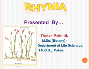
- 1. Presented By… Thakor Maitri M. M.Sc. (Botany) Department of Life Sciences, H.N.G.U., Patan. 1
- 2. CONTENT Introduction classification of Rhynia External features of the plant Internal features of the plant Sporangia of Rhynia Gametophyte stage in Rhynia Life cycle 2
- 3. INTRODUCTION The fossils of the genus Rhynia were discovered by Kidston and Lang in 1917,1921 from the Rhynie locality (chert) of Aberdeenshire in Scotland. ( in middle Devonian Era) 3
- 4. According to Parihar (1996) the chert deposites at Rhynie, originally thought to be of middle Devonian are now believed to be lower Devonian (Late Siegenian or Emsian epoch – about 390 to 374 million years ago. The specimens are so well preserved that they give detailed information about the form and structure of this very primitive vascular plants. Rhynia is a single- species genus of Devonian Vascular plants. 4
- 5. CLASSIFICATION Division : Pteridophyta Sub Division : Psilophyta Class : Psilophytopsida Order : Psilophytales Family : Rhyniaceae Genus : Rhynia 5
- 6. EXTERNAL FEATURES OF THE PLANT Kidston and Lang(1951), thought and supported by estimony that in those times the plants grew in swampy marshes near the volcanoes. The genus is named after the locality and possess two species : (1)Rhynia gwynne vaughani (2)Rhynia major. R. major & R. gwynne vaughani were herbaceous plants. R. major was larger than R. gwynne vaughani. 6
- 7. The plant was differentiated into horizontally cree- ping rhizome & an upright branched shoot without - leaves. There were rootless.7
- 8. The aerial stem of R. major were around 50 cms high and 1.5 mm to 6 mm in diameter , whereas, the corresponding structures of R. gwynne vaughani were 20 cms in height & 1 to 3 mm in diameter. The aerial branches were cylindrical, naked, leafless, dichotomously forked & tapering at their apices. The aerial branches end in tapering vegetative apices or bore pear – shaped terminal sporangia. 8
- 9. INTERNAL FEATURES OF THE PLANTS The internal organization (anato-my) of the rhizome as well as of the aerial stem was practically similar in both species of Rhynia except that some of the slenderest twigs of R. gwynne vaughani were destitute of any vascular structure. In the anatomy of the aerial stem are as followed :9
- 10. Epidermis : ♣ It was the outermost single- layered envelope covered by a thick layer of cuticle. The epidermis of the aerial shoot was interrupted by stomata. 10
- 11. ♣ Each stomata have two guard cells, therefore, the rhizome didn’t contain any stomata. The rhizome possessed unicellular rhizoids on it’s surface. Cortex : ☻The epidermis was followed by a well organised and make broad zone at cortex. ☻The cortex is differentiated into two zones. (a)The outer cortex(hypodermis) (b)The inner cortex 11
- 12. (a) Outer cortex : ☻ The outer cortex consisted of 1-4 cells layer of compactly arranged polygonal paranchy-matus cells without any inter-cellular spaces. ♣ This region perhaps represents hypodermis. (b) Inner cortex : ☻ Inner to the hypodermis, there was broad zone of inner cortex. 12
- 13. ♣ The inner cortex was composed of spherical parenchymatous cells with large intercellular spaces which maintained continuity with the outer atmosphere through the stomata present in the epidermis. ♣ This indicates that the inner cortex was the chief photosynthetic region. (c) Stele / Central cylinder : ☻ A protostele was present in the central part of the axis as well as rhizome. 13
- 14. ♣ The xylem was surrounded by the phloem. ♠ The xylem was composed of only tracheids with annular or spiral thickening. ♣ The phloem was represented by 4 - 5 layers of thin walled elongated cells with oblique end walls. 14
- 15. THE REPRODUCTIVE STRUCTURE (THE SPORANGIA ) Sporangia were borne singly on the apices of some of the aerial branches. They were oval / slightly cylindri-cal / club / elongated shaped, with a diameter slightly greater than that of the subtending branch tip. The sporangia of R. major ; Length: 12 mm , Breath: 1.4 mm Spore : 65µ in diameter. 15
- 16. The sporangia of R. gwynne vaughani ; Length: 4 mm , Breath: 1.4 mm , Spore : 40µ in diameter. Their wall is three layered; (a) Outer epi- Dermal. (b)Middle three cell rows thick layer & (c) an inner layer . ♣The outer layer heavily cutinized. 16
- 17. GAMETOPHYTE STAGE OF RHYNIA Within the same Rhynie chert beds were some germinating spores which show multicellular structure developing at the end of germ tube. This was the indication of the presence of gametophyte in Rhynia. Merker (1961) opined that the underground creeping parts of Rhynia is the gametophyte but not the rhizome. ♣ As sex organs were not presents in these organs, the gametophyte nature because speculative only. 17
- 18. Pant (1960) regarded the small sized R. gwynne vaughani to the gametophytic part of R. major. Puri (1961) is of opinion of the plants described as sporophyte may be gametophytes. 18
- 19. LIFE CYCLE OF RHYNIA 19
- 20. REFERENCES [1] Botany For Degree Students – Pteridophyta ♣ Edition : 2006 ♣ Authors : P.C Vashishta, A.K. Sinha, Dr. Anil kumar. [2] Pteridophyta ♣ Edition : 2015 ♣ Author : S. K. Singh. [3] A text book of Botany ♣ Edition : Volume – 1 ♣ Author : N. C. Kumar. [4] WWW.Slideshsre.net 20
- 21. 21