. The structure and development of mosses and ferns (Archegoniatae). Plant morphology; Mosses; Ferns. THE JUNGERMANNIALES 77 taking place about the same time as the first division in the archegonial cell (Fig. 31, B). The divisions in the latter are the same as in Riccia, and the general structure of the arche- gonium ofifers no noteworthy peculiarities. The number of neck canal cells is small, probably never exceeding four, and in this respect recalls again Riccia. The central cell is relatively large, and the ventral canal cell often nearly as large as the egg. As the archegonium develops, i
Image details
Contributor:
Central Historic Books / Alamy Stock PhotoImage ID:
PG05WKFile size:
7.1 MB (228.5 KB Compressed download)Releases:
Model - no | Property - noDo I need a release?Dimensions:
1733 x 1442 px | 29.3 x 24.4 cm | 11.6 x 9.6 inches | 150dpiMore information:
This image is a public domain image, which means either that copyright has expired in the image or the copyright holder has waived their copyright. Alamy charges you a fee for access to the high resolution copy of the image.
This image could have imperfections as it’s either historical or reportage.
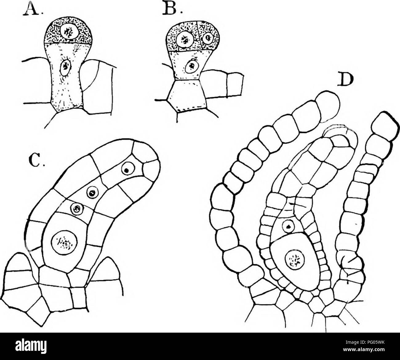
. The structure and development of mosses and ferns (Archegoniatae). Plant morphology; Mosses; Ferns. THE JUNGERMANNIALES 77 taking place about the same time as the first division in the archegonial cell (Fig. 31, B). The divisions in the latter are the same as in Riccia, and the general structure of the arche- gonium ofifers no noteworthy peculiarities. The number of neck canal cells is small, probably never exceeding four, and in this respect recalls again Riccia. The central cell is relatively large, and the ventral canal cell often nearly as large as the egg. As the archegonium develops, its growth is stronger on the posterior side, and it thus curves forward. At first the young archegonium projects free above the surface, but pres-. FlG. 31.—Spharocarpus sp. (?). Development of the archegonium. A-C, Longi- tudinal sections, X6oo; D, X300. ently an envelope is formed about it exactly as in Riccia, but arising at a later stage. After this has begun to form, its growth is very rapid, and it soon overtakes the archegonium and grows beyond it, and finally forms a vesicular body, plainly visible to the naked eye, at the bottom of which the arche- gonium lies. The formation of this involucre is quite inde- pendent of the fertilisation of the archegonium, and as these peculiar vesicles cover completely the whole dorsal surface of the plant, they give it a most characteristic appearance. Usu- ally each archegonium has its own envelope, but Leitgeb ((7), . Please note that these images are extracted from scanned page images that may have been digitally enhanced for readability - coloration and appearance of these illustrations may not perfectly resemble the original work.. Campbell, Douglas Houghton, 1859-1953. New York, The Macmillan Company;